 E-submission
Search
- Page Path
- HOME > Search
Review Article
- Central nervous system tumors with BCOR internal tandem duplications: a systematic review of clinical, radiological, and pathological features in 69 cases
- Ji Young Lee, Sung Sun Kim, Hee Jo Baek, Tae-Young Jung, Kyung-Sub Moon, Jae-Hyuk Lee, Kyung-Hwa Lee
- J Pathol Transl Med. 2025;59(5):273-280. Published online September 1, 2025
- DOI: https://doi.org/10.4132/jptm.2025.07.23
- 5,555 View
- 213 Download
- 1 Web of Science
- 1 Crossref
-
 Abstract
 PDF
PDF Supplementary Material
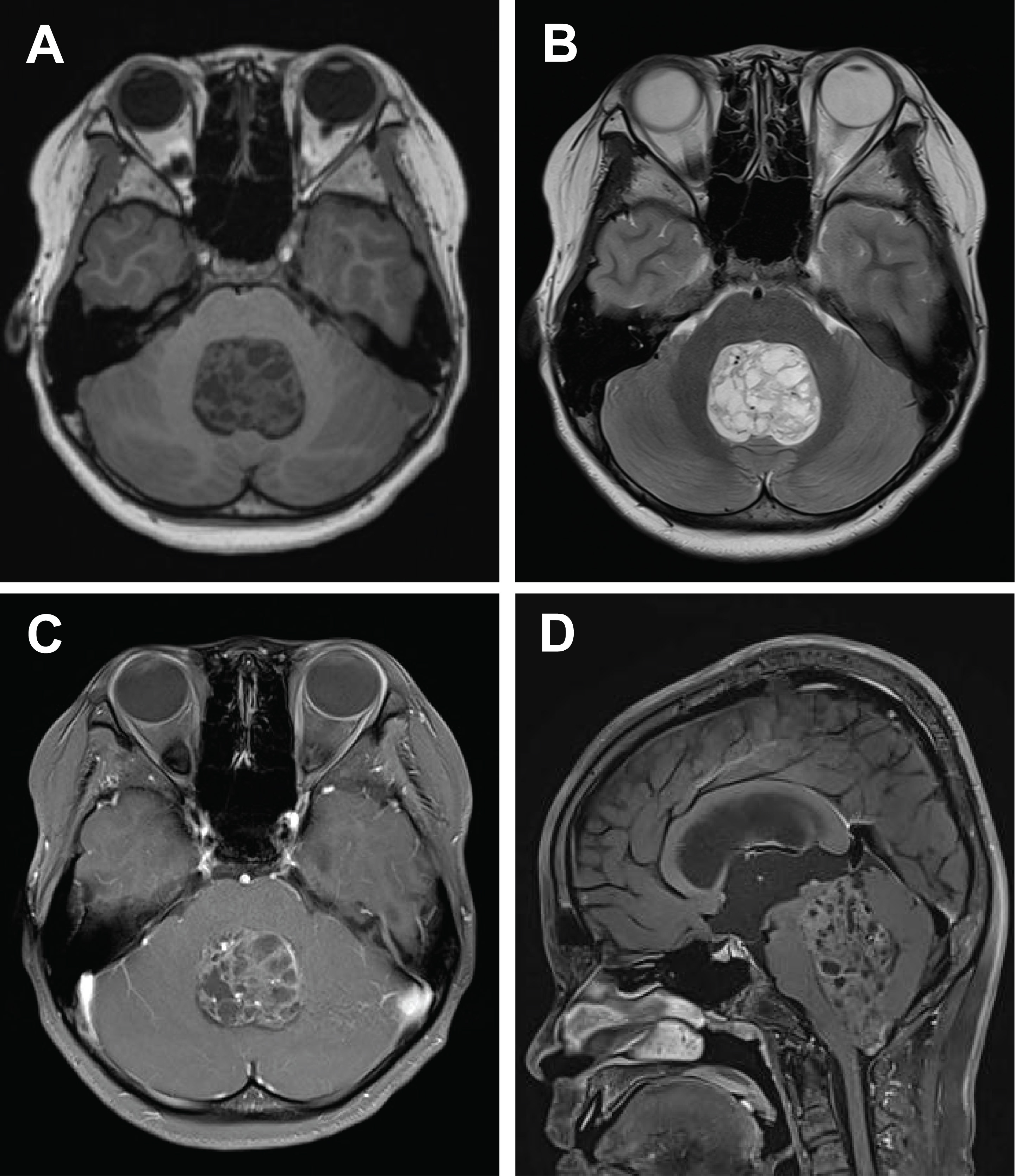
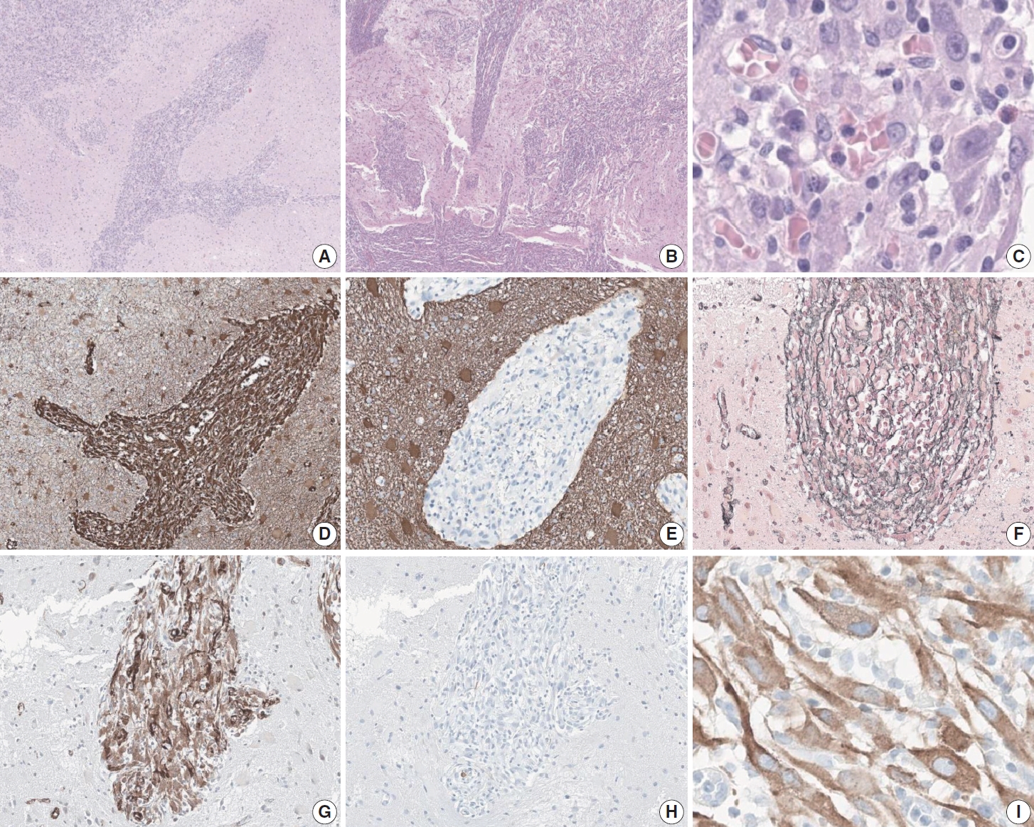
 - Central nervous system tumors with BCL6 corepressor (BCOR) internal tandem duplications (ITDs) constitute a rare, recently characterized pediatric neoplasm with distinct molecular and histopathological features. To date, 69 cases have been documented in the literature, including our institutional case. These neoplasms predominantly occur in young children, with the cerebellum representing the most frequent anatomical location. Radiologically, these tumors present as large, well-circumscribed masses frequently demonstrating necrosis, hemorrhage, and heterogeneous enhancement. Histologically, they are characterized by a monomorphic cellular population featuring ependymoma-like perivascular pseudorosettes, myxoid stroma, and elevated mitotic activity. Immunohistochemically, these tumors exhibit sparse glial fibrillary acidic protein expression while consistently demonstrating positive staining for vimentin and CD56. The defining molecular hallmark is a heterozygous ITD within exon 15 of the BCOR gene, with insertions ranging from 9 to 42 amino acids in length. BCOR immunohistochemistry reveals nuclear positivity in 97.9% of examined cases, although this finding is not pathognomonic for BCOR ITDs. This comprehensive review synthesizes data from all published cases of this novel tumor entity, providing a detailed analysis of clinical presentation, neuroimaging findings, histopathological features with differential diagnostic considerations, therapeutic approaches, and prognostic outcomes.
-
Citations
Citations to this article as recorded by
- Guidance for the Diagnosis and Treatment of Rare Embryonal and
Sarcomatous Brain Tumors—a Report from the Central Nervous System-International
Registry for Rare Embryonal and Sarcomatous Tumors German Society of Ped
Pascal Johann, Dominik Sturm, Rolf Kortmann, Brigitte Bison, David Capper, Ahmed El Damaty, Lars Behrens, Selma Manea, Johannes Gojo, Michael Frühwald, Ulrich-Wilhelm Thomale, Martin U. Schuhmann, Rudolf Schwarz, Anna Tietze, Matthias Wagner, Beate Timmer
Klinische Pädiatrie.2026;[Epub] CrossRef
- Guidance for the Diagnosis and Treatment of Rare Embryonal and
Sarcomatous Brain Tumors—a Report from the Central Nervous System-International
Registry for Rare Embryonal and Sarcomatous Tumors German Society of Ped
Case Study
- Primary epithelioid inflammatory myofibroblastic sarcoma of the brain with EML4::ALK fusion mimicking intra-axial glioma: a case report and brief literature review
- Eric Eunshik Kim, Chul-Kee Park, Koung Mi Kang, Yoonjin Kwak, Sung-Hye Park, Jae-Kyung Won
- J Pathol Transl Med. 2024;58(3):141-145. Published online May 14, 2024
- DOI: https://doi.org/10.4132/jptm.2024.04.12
- 5,973 View
- 215 Download
- 2 Web of Science
- 2 Crossref
-
Abstract
PDF
- An aggressive subtype of inflammatory myofibroblastic tumor, epithelioid inflammatory myofibroblastic sarcoma occurs primarily inside the abdominal cavity, followed by a pulmonary localization. Most harbor anaplastic lymphoma kinase (ALK) gene rearrangements, with RANBP2 and RRBP1 among the well-documented fusion partners. We report the second case of primary epithelioid inflammatory myofibroblastic sarcoma of the brain, with a well-known EML4::ALK fusion. The case is notable for its intra-axial presentation that clinico-radiologically mimicked glioma.
-
Citations
Citations to this article as recorded by
Original Article
- Single-center study on clinicopathological and typical molecular pathologic features of metastatic brain tumor
- Su Hwa Kim, Young Suk Lee, Sung Hak Lee, Yeoun Eun Sung, Ahwon Lee, Jun Kang, Jae-Sung Park, Sin Soo Jeun, Youn Soo Lee
- J Pathol Transl Med. 2023;57(4):217-231. Published online July 11, 2023
- DOI: https://doi.org/10.4132/jptm.2023.06.10
- 7,109 View
- 176 Download
- 1 Crossref
-
Abstract
PDF
- Background
The metastatic brain tumor is the most common brain tumor. The aim of this study was to demonstrate the clinicopathological and molecular pathologic features of brain metastases (BM).
Methods
A total of 269 patients were diagnosed with BM through surgical resection at Seoul St. Mary’s Hospital from January 2010 to March 2020. We reviewed the clinicopathological features and molecular status of primary and metastatic brain tissues using immunohistochemistry and molecular pathology results.
Results
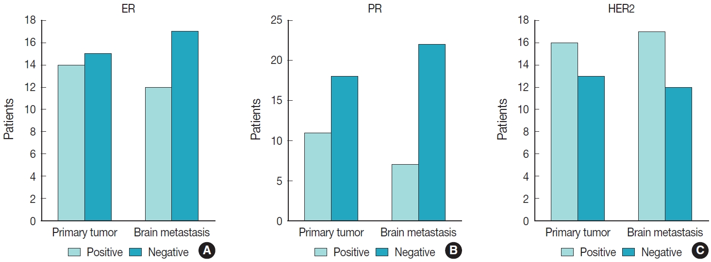
Among 269 patients, 139 males and 130 females were included. The median age of primary tumor was 58 years (range, 13 to 87 years) and 86 patients (32.0%) had BM at initial presentation. Median BM free interval was 28.0 months (range, 1 to 286 months). The most frequent primary site was lung 46.5% (125/269), and followed by breast 15.6% (42/269), colorectum 10.0% (27/269). Epidermal growth factor receptor (EGFR) mutation was found in 50.8% (32/63) and 58.0% (40/69) of lung primary and BM, respectively. In both breast primary and breast cancer with BM, luminal B was the most frequent subtype at 37.9% (11/29) and 42.9% (18/42), respectively, followed by human epidermal growth factor receptor 2 with 31.0% (9/29) and 33.3% (14/42). Triple-negative was 20.7% (6/29) and 16.7% (7/42), and luminal A was 10.3% (3/29) and 7.1% (3/42) of breast primary and BM, respectively. In colorectal primary and colorectal cancer with BM, KRAS mutation was found in 76.9% (10/13) and 66.7% (2/3), respectively.
Conclusions
We report the clinicopathological and molecular pathologic features of BM that can provide useful information for understanding the pathogenesis of metastasis and for clinical trials based on the tumor’s molecular pathology. -
Citations
Citations to this article as recorded by- Colorectal cancer metastasis to the brain: A scoping review of incidence, treatment, and outcomes
Hunter J Hutchinson, Melanie Gonzalez, Diana Feier, Colin E Welch, Brandon Lucke-Wold
World Journal of Gastrointestinal Pathophysiology.2025;[Epub] CrossRef
- Colorectal cancer metastasis to the brain: A scoping review of incidence, treatment, and outcomes
Reviews
- Neuropathologic features of central nervous system hemangioblastoma
- Rebecca A. Yoda, Patrick J. Cimino
- J Pathol Transl Med. 2022;56(3):115-125. Published online May 3, 2022
- DOI: https://doi.org/10.4132/jptm.2022.04.13
- 19,571 View
- 388 Download
- 24 Web of Science
- 30 Crossref
-
Abstract
PDF
- Hemangioblastoma is a benign, highly vascularized neoplasm of the central nervous system (CNS). This tumor is associated with loss of function of the VHL gene and demonstrates frequent occurrence in von Hippel-Lindau (VHL) disease. While this entity is designated CNS World Health Organization grade 1, due to its predilection for the cerebellum, brainstem, and spinal cord, it is still an important cause of morbidity and mortality in affected patients. Recognition and accurate diagnosis of hemangioblastoma is essential for the practice of surgical neuropathology. Other CNS neoplasms, including several tumors associated with VHL disease, may present as histologic mimics, making diagnosis challenging. We outline key clinical and radiologic features, pathophysiology, treatment modalities, and prognostic information for hemangioblastoma, and provide a thorough review of the gross, microscopic, immunophenotypic, and molecular features used to guide diagnosis.
-
Citations
Citations to this article as recorded by- Renal hemangioblastoma and renal cell carcinoma with fibromyomatous stroma and hemangioblastoma-like areas belong to the spectrum of one entity
Kiril Trpkov, Norel Salut, Inmaculada Ribera-Cortada, Elías Tasso Xipell, Isabel Trias Puigsureda, Asli Yilmaz, Arjumand Riyaz Husain, Erik Nohr, Adrian Box, Farshid Siadat, Katherina Baranova, Rola M. Saleeb, Robert Stoehr, Arndt Hartmann, Abbas Agaimy
Virchows Archiv.2026; 488(4): 767. CrossRef - Hemangioblastoma of the Kidney—A Comprehensive Clinical, Pathological, and Genetic Analysis of Four Cases
Boglárka Pósfai, Alex Jenei, Gertrúd Forika, Attila Fintha, Zoltán Sápi, Áron Somorácz, Borbála Dénes, Ferenc Salamon, Kornélia Veronika Eizler, Nándor Giba, Dávid Semjén, Ildikó Illyés, Kristóf Attila Kovács, Gyöngyi Munkácsy, János Papp, Fanni Sánta, He
APMIS.2026;[Epub] CrossRef - Peritumoral Cystic Meningioma With Diagnostic Challenges: Two Case Reports and a Literature Review
Wei-Chih Chen, Zi-Jie Lin, Lam Chee-Tat
Cureus.2026;[Epub] CrossRef - Belzutifan-induced tumor regression in sporadic hemangioblastoma: a case report and literature review
Rebekka E. Hooks, Niket Yadav, Mark Willy L. Mondia, Georgios Mantziaris, Alaa Saleh, Anna Vi Jones, Matthew McCord, Melike Mut, Ashok R. Asthagiri, Benjamin W. Purow
Journal of Neuro-Oncology.2026;[Epub] CrossRef - Unusual immunohistochemical profiles in hemangioblastomas and their relevance in differential diagnosis: A comprehensive study of 112 cases
Jiri Soukup, Marie Novakova, Jan Hojny, Marketa Trnkova, Martin Syrucek, Tomas Jirasek, Patricie Delongova, Ales Kohout, Tomas Vebr, Jan Sroubek, Alena Sejkorova, Radim Lipina, Radim Brabec, Tomas Cesak, David Netuka
Journal of Neuropathology & Experimental Neurology.2026;[Epub] CrossRef - Pathological features and clinical outcomes of hemangioblastoma in the spinal cord of three dogs
Ryo SAITO, James K CHAMBERS, Kio YOSHIDA, Yutaro NAKAYAMA, Yukiko NAKANO, Kosuke HAII, Yumiko KAGAWA, Kazuyuki UCHIDA
Journal of Veterinary Medical Science.2026; 88(4): 568. CrossRef - Monitoring and treatment patterns of von Hippel-Lindau disease-associated central nervous system hemangioblastomas
Eric Jonasch, Yan Song, Jonathan Freimark, Manasi Mohan, James Signorovitch, Murali Sundaram
Hereditary Cancer in Clinical Practice.2026;[Epub] CrossRef - Multimodal Management of Spinal Cord Hemangioblastomas: A Comprehensive Review
Francisco Alfredo Call-Orellana, Juan Pablo Zuluaga-Garcia, Maria Alejandra Sierra, Mariana Zuluaga-Garcia, Esteban Ramirez-Ferrer, Alejandro Bugarini
Therapeutics.2026; 3(2): 12. CrossRef - FLOW800−guided microsurgical resection of a brainstem hemangioblastoma with AVM−like features: a case report and technical note
Ying Yang, Chunfeng Pan, Chuan He, Zhaobin Chen, Zhaoyi Quan, Gang Cao
Frontiers in Oncology.2026;[Epub] CrossRef - Immunohistochemical Expression of PAX8 in Central Nervous System Hemangioblastomas: A Potential Diagnostic Pitfall for Neuropathologists
Giuseppe Broggi, Jessica Farina, Valeria Barresi, Francesco Certo, Giuseppe Maria Vincenzo Barbagallo, Gaetano Magro, Rosario Caltabiano
Applied Immunohistochemistry & Molecular Morphology.2025; 33(3): 160. CrossRef - Endolymphatic Sac Tumor. Post-Radiosurgery Evaluation Using Time-Resolved Imaging of Contrast Kinetics MR Angiography
Antonella Blandino, Allegra Romano, Chiara Filippi, Sofia Pizzolante, Andrea Romano, Giulia Moltoni, Edoardo Covelli, Maurizio Barbara, Alessandro Bozzao
Ear, Nose & Throat Journal.2025;[Epub] CrossRef - Stereotactic radiosurgery in the management of central nervous system hemangioblastomas: a systematic review and meta-analysis
Amirhossein Zare, Amirhessam Zare, Alireza Soltani Khaboushan, Bardia Hajikarimloo, Jason P. Sheehan
Neurosurgical Review.2025;[Epub] CrossRef - Cerebellar medullary cistern hemangioblastoma
Dahai Cao, Qiang Zhang
Asian Journal of Surgery.2025; 48(9): 5843. CrossRef - Navigating rare vascular brain tumors: A retrospective observational study
Sana Ahuja, Dipanker S Mankotia, Naveen Kumar, Vyomika Teckchandani, Sufian Zaheer
Cancer Research, Statistics, and Treatment.2025; 8(2): 92. CrossRef - A potential new entity pending further validation of pulmonary primary interstitial Tumor: Lymphangioleiomyomatosis-like
Lingyu Zhao, Xiaochen Shen, Yun Niu, Huang Chen, Dingrong Zhong
Respiratory Medicine Case Reports.2025; 57: 102241. CrossRef - Renal cell carcinoma with fibromyomatous stroma (RCC FMS) and with hemangioblastoma‐like areas is part of the RCC FMS spectrum in patients with tuberous sclerosis complex
Katherina Baranova, Jacob A Houpt, Deaglan Arnold, Andrew A House, Laura Lockau, Lindsay Ninivirta, Stephen Pautler, Haiying Chen, Madeleine Moussa, Rola Saleeb, Jose A Gomez, Asli Yilmaz, Farshid Siadat, Adrian Box, Douglas J Mahoney, Franz J Zemp, Manal
Histopathology.2025; 87(5): 687. CrossRef - Primary hemangioblastoma of rectum: a rare case report and review of literature
Aiping Zheng, Shaojuan Zhang, Qiang Ma, Wenxu Yang, Hualiang Xiao, Xinyu Liang
Journal of Cancer Research and Clinical Oncology.2025;[Epub] CrossRef - Cerebellar Hemangioblastoma Resection Complicated by Postoperative Vasogenic Edema in the Setting of Concurrent Immunotherapy Treatment
Aashka Sheth, Nicholas Dietz, Andrea Becerril-Gaitan, Rahim Kasem, Akshitkumar Mistry, Brian J Williams, Dale Ding, Isaac Abecassis
Cureus.2025;[Epub] CrossRef - Familial Von Hippel–Lindau Disease: A Case Series of Cerebral Hemangioblastomas with MRI, Histopathological, and Genetic Correlations
Claudiu Matei, Ioana Boeras, Dan Orga Dumitriu, Cosmin Mutu, Adriana Popescu, Mihai Gabriel Cucu, Alexandru Calotă-Dobrescu, Bogdan Fetica, Diter Atasie
Life.2025; 15(11): 1649. CrossRef - Characterization of spinal hemangioblastomas in patients with and without von Hippel-Lindau, and YAP expression
Ana-Laura Calderón-Garcidueñas, Steven-Andrés Piña-Ballantyne, Eunice-Jazmín Espinosa-Aguilar, Rebeca de Jesús Ramos-Sánchez
Revista Española de Patología.2024; 57(3): 160. CrossRef - Patients With Hemangioblastoma: Mood Disorders and Sleep Quality
Ali Riazi, Yaser Emaeillou, Nima Najafi, Mohammad Hoseinimanesh, Mohammad Ibrahim Ashkaran, Donya Sheibani Tehrani
Brain Tumor Research and Treatment.2024; 12(2): 87. CrossRef - Radiosurgically Treated Recurrent Cerebellar Hemangioblastoma: A Case Report and Literature Review
François Fabi, Ève Chamberland, Myreille D’Astous, Karine Michaud, Martin Côté, Isabelle Thibault
Current Oncology.2024; 31(7): 3968. CrossRef - Dual manifestations: spinal and cerebellar hemangioblastomas indicative of von Hippel-Lindau syndrome
Nurhuda Hendra Setyawan, Rachmat Andi Hartanto, Rusdy Ghazali Malueka, Ery Kus Dwianingsih, Dito Pondra Dharma
Radiology Case Reports.2024; 19(11): 5000. CrossRef - Phenotypic and Genotypic Features of a Chinese Cohort with Retinal Hemangioblastoma
Liqin Gao, Feng Zhang, J. Fielding Hejtmancik, Xiaodong Jiao, Liyun Jia, Xiaoyan Peng, Kai Ma, Qian Li
Genes.2024; 15(9): 1192. CrossRef - Hemangioblastoma Incidentally Discovered at CT Scan in Bamako: About a Case
Traore Ousmane, N’Diaye Mamadou, Dembélé Mamadou, Dembélé Adama, Diakité Siaka, Sidibé Mansa Drissa, Camara Nagnoumague, Keita Adama Diaman
Open Journal of Medical Imaging.2024; 14(03): 123. CrossRef - Case report: Hemangioblastoma in the brainstem of a dog
Kirsten Landsgaard, Samantha St. Jean, Stephanie Lovell, Jonathan Levine, Christine Gremillion, Brian Summers, Raquel R. Rech
Frontiers in Veterinary Science.2023;[Epub] CrossRef - Intramedullary hemangioblastoma of the thoracic cord with a microsurgical approach: A case report and literature review
Eduardo Cattapan Piovesan, Werner Petry Silva, Adroaldo Baseggio Mallmann, Felipe Severo Lanzini, Bruna Zanatta de Freitas, Francisco Costa Beber Lemanski, Charles André Carazzo
Surgical Neurology International.2023; 14: 137. CrossRef - Secondary Holocord Syringomyelia Associated With Spinal Hemangioblastoma in a 29-Year-Old Female
Eric Chun-Pu Chu, Edouard Sabourdy, Benjamin Cheong
Cureus.2023;[Epub] CrossRef - Belzutifan in adults with VHL-associated central nervous system hemangioblastoma: a single-center experience
Bryan J. Neth, Mason J. Webb, Jessica White, Joon H. Uhm, Pavel N. Pichurin, Ugur Sener
Journal of Neuro-Oncology.2023; 164(1): 239. CrossRef - Resection of Intramedullary Hemangioblastoma: Timing of Surgery and Its Impact on Neurological Outcome and Quality of Life
Michael Schwake, Sarah Ricchizzi, Sophia Krahwinkel, Emanuele Maragno, Stephanie Schipmann, Walter Stummer, Marco Gallus, Markus Holling
Medicina.2023; 59(9): 1611. CrossRef
- Renal hemangioblastoma and renal cell carcinoma with fibromyomatous stroma and hemangioblastoma-like areas belong to the spectrum of one entity
- Molecular Testing of Brain Tumor
- Sung-Hye Park, Jaekyung Won, Seong-Ik Kim, Yujin Lee, Chul-Kee Park, Seung-Ki Kim, Seung-Hong Choi
- J Pathol Transl Med. 2017;51(3):205-223. Published online May 12, 2017
- DOI: https://doi.org/10.4132/jptm.2017.03.08
- 38,890 View
- 1,183 Download
- 43 Web of Science
- 50 Crossref
-
Abstract
PDF
- The World Health Organization (WHO) classification of central nervous system (CNS) tumors was revised in 2016 with a basis on the integrated diagnosis of molecular genetics. We herein provide the guidelines for using molecular genetic tests in routine pathological practice for an accurate diagnosis and appropriate management. While astrocytomas and IDH-mutant (secondary) glioblastomas are characterized by the mutational status of IDH, TP53, and ATRX, oligodendrogliomas have a 1p/19q codeletion and mutations in IDH, CIC, FUBP1, and the promoter region of telomerase reverse transcriptase (TERTp). IDH-wildtype (primary) glioblastomas typically lack mutations in IDH, but are characterized by copy number variations of EGFR, PTEN, CDKN2A/B, PDGFRA, and NF1 as well as mutations of TERTp. High-grade pediatric gliomas differ from those of adult gliomas, consisting of mutations in H3F3A, ATRX, and DAXX, but not in IDH genes. In contrast, well-circumscribed low-grade neuroepithelial tumors in children, such as pilocytic astrocytoma, pleomorphic xanthoastrocytoma, and ganglioglioma, often have mutations or activating rearrangements in the BRAF, FGFR1, and MYB genes. Other CNS tumors, such as ependymomas, neuronal and glioneuronal tumors, embryonal tumors, meningothelial, and other mesenchymal tumors have important genetic alterations, many of which are diagnostic, prognostic, and predictive markers and therapeutic targets. Therefore, the neuropathological evaluation of brain tumors is increasingly dependent on molecular genetic tests for proper classification, prediction of biological behavior and patient management. Identifying these gene abnormalities requires cost-effective and high-throughput testing, such as next-generation sequencing. Overall, this paper reviews the global guidelines and diagnostic algorithms for molecular genetic testing of brain tumors.
-
Citations
Citations to this article as recorded by- Low-grade Epilepsy-associated Tumors and Other Acquired Causes of Epilepsy
Horst Urbach, Soroush Doostkam
Neuroimaging Clinics of North America.2026; 36(2): 273. CrossRef - Barriers to care to pediatric brain tumors in low- and middle-income countries: A proposal for a critical framework
Savi Agarwal, Jorge Lazareff
Surgical Neurology International.2026; 17: 154. CrossRef - Radiomics and deep learning models for predicting glioma p53 status: A diagnostic accuracy systematic review and meta-analysis of magnetic resonance imaging studies
Amir Mahmoud Ahmadzadeh, Mohammad Amin Ashoobi, Nima Broomand Lomer, Mahsa Vatanparast, Benyamin Gheiji, Danial Elyassirad, Shahriar Faghani
Clinical Imaging.2026; 135: 110817. CrossRef - Brain Cancer: Molecular Alterations and Emerging Trends in Neuropharmacology
Beata Leskova, Ilaria D’Agostino, Simona Mattova, Nicol Urbanska, Alzbeta Blicharova, Patrik Simko, Aylin Toplu, Muhammet Karaman, Terezia Kiskova-Simkova
International Journal of Molecular Sciences.2026; 27(11): 4880. CrossRef - Navigating rare vascular brain tumors: A retrospective observational study
Sana Ahuja, Dipanker S Mankotia, Naveen Kumar, Vyomika Teckchandani, Sufian Zaheer
Cancer Research, Statistics, and Treatment.2025; 8(2): 92. CrossRef - Author’s reply to Dagar and Mallick
Sana Ahuja, Sufian Zaheer
Cancer Research, Statistics, and Treatment.2025; 8(3): 245. CrossRef - The Clinical Utility of Cell-Free DNA in Brain Tumor Management: A Comprehensive Review
Qama Abuhassan, Hanan Hassan Ahmed, Radhwan Abdul Kareem, Soumya V. Menon, Priya Priyadarshini Nayak, J. Bethanney Janney, Vimal Arora, Aashna Sinha, Saif Aldeen Jaber, Hayder Naji Sameer, Ahmed Yaseen, Zainab H. Athab, Mohaned Adil
Journal of Molecular Neuroscience.2025;[Epub] CrossRef - PTEN regulates expression of its pseudogene in glioblastoma cells in DNA methylation-dependent manner
Tatyana F. Kovalenko, Bhupender Yadav, Ksenia S. Anufrieva, Tatyana D. Larionova, Tatiana E. Aksinina, Yaroslav A. Latyshev, Soniya Bastola, Michail I. Shakhparonov, Amit Kumar Pandey, Marat S. Pavlyukov
Biochimie.2024; 219: 74. CrossRef - CDKN2A/B deletion in IDH-mutant astrocytomas: An evaluation by Fluorescence in-situ hybridization
Manali Ranade, Sridhar Epari, Omshree Shetty, Sandeep Dhanavade, Sheetal Chavan, Ayushi Sahay, Arpita Sahu, Prakash Shetty, Aliasgar Moiyadi, Vikash Singh, Archya Dasgupta, Abhishek Chatterjee, Sadhana Kannan, Tejpal Gupta
Journal of Neuro-Oncology.2024; 167(1): 189. CrossRef - Central neurocytoma exhibits radial glial cell signatures with FGFR3 hypomethylation and overexpression
Yeajina Lee, Tamrin Chowdhury, Sojin Kim, Hyeon Jong Yu, Kyung-Min Kim, Ho Kang, Min-Sung Kim, Jin Wook Kim, Yong-Hwy Kim, So Young Ji, Kihwan Hwang, Jung Ho Han, Jinha Hwang, Seong-Keun Yoo, Kyu Sang Lee, Gheeyoung Choe, Jae-Kyung Won, Sung-Hye Park, Yon
Experimental & Molecular Medicine.2024; 56(4): 975. CrossRef - Liquid biopsy in brain tumors: Potential for impactful clinical applications
Tania Eid, Lina Ghandour, Joseph Abi Ghanem, Hazem Assi, Rami Mahfouz
Human Gene.2024; 42: 201333. CrossRef - Diagnostic challenges in complicated case of glioblastoma
Tatiana Aghova, Halka Lhotska, Libuse Lizcova, Karla Svobodova, Lucie Hodanova, Karolina Janeckova, Kim Vucinic, Martin Gregor, Dora Konecna, Filip Kramar, Jiri Soukup, David Netuka, Zuzana Zemanova
Pathology and Oncology Research.2024;[Epub] CrossRef - Drugging Hijacked Kinase Pathways in Pediatric Oncology: Opportunities and Current Scenario
Marina Ferreira Candido, Mariana Medeiros, Luciana Chain Veronez, David Bastos, Karla Laissa Oliveira, Julia Alejandra Pezuk, Elvis Terci Valera, María Sol Brassesco
Pharmaceutics.2023; 15(2): 664. CrossRef - BrainBase: a curated knowledgebase for brain diseases
Lin Liu, Yang Zhang, Guangyi Niu, Qianpeng Li, Zhao Li, Tongtong Zhu, Changrui Feng, Xiaonan Liu, Yuansheng Zhang, Tianyi Xu, Ruru Chen, Xufei Teng, Rongqin Zhang, Dong Zou, Lina Ma, Zhang Zhang
Nucleic Acids Research.2022; 50(D1): D1131. CrossRef - A heat shock protein 90 inhibitor reduces oncoprotein expression and induces cell death in heterogeneous glioblastoma cells with EGFR, PDGFRA, CDK4, and NF1 aberrations
Kuan-Ta Ho, Pei-Fan Chen, Jian-Ying Chuang, Po-Wu Gean, Yuan-Shuo Hsueh
Life Sciences.2022; 288: 120176. CrossRef - Bicentric validation of the navigated transcranial magnetic stimulation motor risk stratification model
Tizian Rosenstock, Levin Häni, Ulrike Grittner, Nicolas Schlinkmann, Meltem Ivren, Heike Schneider, Andreas Raabe, Peter Vajkoczy, Kathleen Seidel, Thomas Picht
Journal of Neurosurgery.2022; 136(4): 1194. CrossRef - Foramen magnum meningioma presented as cervical myelopathy in a pregnant COVID-19 patient: A case report
Olivia Josephine Wijaya, Djohan Ardiansyah
Annals of Medicine and Surgery.2022; 77: 103647. CrossRef - The telomere maintenance mechanism spectrum and its dynamics in gliomas
Sojin Kim, Tamrin Chowdhury, Hyeon Jong Yu, Jee Ye Kahng, Chae Eun Lee, Seung Ah. Choi, Kyung-Min Kim, Ho Kang, Joo Ho Lee, Soon-Tae Lee, Jae-Kyung Won, Kyung Hyun Kim, Min-Sung Kim, Ji Yeoun Lee, Jin Wook Kim, Yong-Hwy Kim, Tae Min Kim, Seung Hong Choi,
Genome Medicine.2022;[Epub] CrossRef - A review of predictive, prognostic and diagnostic biomarkers for brain tumours: towards personalised and targeted cancer therapy
Ernest Osei, Pascale Walters, Olivia Masella, Quinton Tennant, Amber Fishwick, Eugenia Dadzie, Anmol Bhangu, Johnson Darko
Journal of Radiotherapy in Practice.2021; 20(1): 83. CrossRef - Use of a novel navigable tubular retractor system in 1826 minimally invasive parafascicular surgery (MIPS) cases involving deep-seated brain tumors, hemorrhages and malformations
Martina M. Cartwright, Penny Sekerak, Joseph Mark, Julian Bailes
Interdisciplinary Neurosurgery.2021; 23: 100919. CrossRef - Disentangling the therapeutic tactics in GBM: From bench to bedside and beyond
S. Daisy Precilla, Shreyas S. Kuduvalli, Anitha Thirugnanasambandhar Sivasubramanian
Cell Biology International.2021; 45(1): 18. CrossRef - Sequencing of a central nervous system tumor demonstrates cancer transmission in an organ transplant
Marie-Claude Gingras, Aniko Sabo, Maria Cardenas, Abbas Rana, Sadhna Dhingra, Qingchang Meng, Jianhong Hu, Donna M Muzny, Harshavardhan Doddapaneni, Lesette Perez, Viktoriya Korchina, Caitlin Nessner, Xiuping Liu, Hsu Chao, John Goss, Richard A Gibbs
Life Science Alliance.2021; 4(9): e202000941. CrossRef - Radiogenomics of Gliomas
Chaitra Badve, Sangam Kanekar
Radiologic Clinics of North America.2021; 59(3): 441. CrossRef - Chemical analysis of the human brain by imaging mass spectrometry
Akhila Ajith, Yeswanth Sthanikam, Shibdas Banerjee
The Analyst.2021; 146(18): 5451. CrossRef - Correlation between Stage and Histopathological Features and Clinical Outcomes in Patients with Glioma Tumors
Andre Lona, Alfansuri Kadri, Irina Kemala Nasution
Open Access Macedonian Journal of Medical Sciences.2021; 9(T3): 262. CrossRef - Pediatric Glioma: An Update of Diagnosis, Biology, and Treatment
Yusuke Funakoshi, Nobuhiro Hata, Daisuke Kuga, Ryusuke Hatae, Yuhei Sangatsuda, Yutaka Fujioka, Kosuke Takigawa, Masahiro Mizoguchi
Cancers.2021; 13(4): 758. CrossRef - Discovery of clinically relevant fusions in pediatric cancer
Stephanie LaHaye, James R. Fitch, Kyle J. Voytovich, Adam C. Herman, Benjamin J. Kelly, Grant E. Lammi, Jeremy A. Arbesfeld, Saranga Wijeratne, Samuel J. Franklin, Kathleen M. Schieffer, Natalie Bir, Sean D. McGrath, Anthony R. Miller, Amy Wetzel, Katheri
BMC Genomics.2021;[Epub] CrossRef - Clinicopathological correlation of glioma patients with respect to immunohistochemistry markers: A prospective study of 115 patients in a Tertiary Care Hospital in North India
Gitanshu Dahuja, Ashok Gupta, Arpita Jindal, Gaurav Jain, Santosh Sharma, Arvind Kumar
Asian Journal of Neurosurgery.2021; 16(04): 732. CrossRef - Role of Extracellular Vesicles in Glioma Progression: Deciphering Cellular Biological Processes to Clinical Applications
Rashmi Rana, Shikha Joon, Kirti Chauhan, Vaishnavi Rathi, Nirmal Kumar Ganguly, Chandni Kumari, Dharmendra Kumar Yadav
Current Topics in Medicinal Chemistry.2021; 21(8): 696. CrossRef - A comprehensive overview on the molecular biology of human glioma: what the clinician needs to know
P. D. Delgado-López, P. Saiz-López, R. Gargini, E. Sola-Vendrell, S. Tejada
Clinical and Translational Oncology.2020; 22(11): 1909. CrossRef - Use of Fluorescence In Situ Hybridization (FISH) in Diagnosis and Tailored Therapies in Solid Tumors
Natalia Magdalena Chrzanowska, Janusz Kowalewski, Marzena Anna Lewandowska
Molecules.2020; 25(8): 1864. CrossRef - From Banding to BAM Files
Adrian M. Dubuc
Surgical Pathology Clinics.2020; 13(2): 343. CrossRef - Mutation profiling of anaplastic ependymoma grade III by Ion Proton next generation DNA sequencing
Ejaz Butt, Sabra Alyami, Tahani Nageeti, Muhammad Saeed, Khalid AlQuthami, Abdellatif Bouazzaoui, Mohammad Athar, Zainularifeen Abduljaleel, Faisal Al-Allaf, Mohiuddin Taher
F1000Research.2020; 8: 613. CrossRef - Assessment of the non-linear optical behavior of cells for discrimination between normal and malignant glial cells
Soraya Emamgholizadeh Minaei, Alireza Ghader, Ali Abbasian Ardakani, Samideh Khoei, Mohammad Hosein Majles Ara
Laser Physics.2020; 30(12): 125601. CrossRef - Brain Tumor Classification in Magnetic Resonance Images Using Deep Learning and Wavelet Transform
Ahmad M. Sarhan
Journal of Biomedical Science and Engineering.2020; 13(06): 102. CrossRef - Mutation profiling of anaplastic ependymoma grade III by Ion Proton next generation DNA sequencing
Muhammad Butt, Sabra Alyami, Tahani Nageeti, Muhammad Saeed, Khalid AlQuthami, Abdellatif Bouazzaoui, Mohammad Athar, Zainularifeen Abduljaleel, Faisal Al-Allaf, Mohiuddin Taher
F1000Research.2019; 8: 613. CrossRef - A Multiplex Quantitative Reverse Transcription Polymerase Chain Reaction Assay for the Detection of KIAA1549–BRAF Fusion Transcripts in Formalin-Fixed Paraffin-Embedded Pilocytic Astrocytomas
David Bret, Valentin Chappuis, Delphine Poncet, François Ducray, Karen Silva, Fabrice Mion, Alexandre Vasiljevic, Carole Ferraro-Peyret, Carmine Mottolese, Pierre Leblond, Mathieu Gabut, Didier Frappaz, Nathalie Streichenberger, David Meyronet, Pierre-Pau
Molecular Diagnosis & Therapy.2019; 23(4): 537. CrossRef - A PTPmu Biomarker is Associated with Increased Survival in Gliomas
Mette L. Johansen, Jason Vincent, Haley Gittleman, Sonya E. L. Craig, Marta Couce, Andrew E. Sloan, Jill S. Barnholtz-Sloan, Susann M. Brady-Kalnay
International Journal of Molecular Sciences.2019; 20(10): 2372. CrossRef -
ATRX Mutations in Pineal Parenchymal Tumors of Intermediate Differentiation
Haydee Martínez, Michelle Nagurney, Zi-Xuan Wang, Charles G Eberhart, Christopher M Heaphy, Mark T Curtis, Fausto J Rodriguez
Journal of Neuropathology & Experimental Neurology.2019; 78(8): 703. CrossRef - The role of fibroblast growth factors and their receptors in gliomas: the mutations involved
Vasiliki Georgiou, Vasiliki Gkretsi
Reviews in the Neurosciences.2019; 30(5): 543. CrossRef - The Role of Next Generation Sequencing in Diagnosis of Brain Tumors: A Review Study
Sadegh Shirian, Yahya Daneshbod, Saranaz Jangjoo, Amir Ghaemi, Arash Goodarzi, Maryam Ghavideldarestani, Ahmad Emadi, Arman Ai, Akbar Ahmadi, Jafar Ai
Archives of Neuroscience.2019;[Epub] CrossRef - Applied Precision Cancer Medicine in Neuro-Oncology
H. Taghizadeh, L. Müllauer, J. Furtner, J. A. Hainfellner, C. Marosi, M. Preusser, G. W. Prager
Scientific Reports.2019;[Epub] CrossRef - Comparative genomic analysis of driver mutations in matched primary and recurrent meningiomas
Joshua Loewenstern, John Rutland, Corey Gill, Hanane Arib, Margaret Pain, Melissa Umphlett, Yayoi Kinoshita, Russell McBride, Michael Donovan, Robert Sebra, Joshua Bederson, Mary Fowkes, Raj Shrivastava
Oncotarget.2019; 10(37): 3506. CrossRef - Next generation DNA sequencing of atypical choroid plexus papilloma of brain: Identification of novel mutations in a female patient by Ion Proton
Mohiuddin Taher, Amal Hassan, Muhammad Saeed, Raid Jastania, Tahani Nageeti, Hisham Alkhalidi, Ghida Dairi, Zainularifeen Abduljaleel, Mohammad Athar, Abdellatif Bouazzaoui, Wafa El‑Bjeirami, Faisal Al‑Allaf
Oncology Letters.2019;[Epub] CrossRef - Pure mechanistic analysis of additive neuroprotective effects between baicalin and jasminoidin in ischemic stroke mice
Peng-qian Wang, Qiong Liu, Wen-juan Xu, Ya-nan Yu, Ying-ying Zhang, Bing Li, Jun Liu, Zhong Wang
Acta Pharmacologica Sinica.2018; 39(6): 961. CrossRef - The Smad4/PTEN Expression Pattern Predicts Clinical Outcomes in Colorectal Adenocarcinoma
Yumin Chung, Young Chan Wi, Yeseul Kim, Seong Sik Bang, Jung-Ho Yang, Kiseok Jang, Kyueng-Whan Min, Seung Sam Paik
Journal of Pathology and Translational Medicine.2018; 52(1): 37. CrossRef - Primary brain tumours in adults
Sarah Lapointe, Arie Perry, Nicholas A Butowski
The Lancet.2018; 392(10145): 432. CrossRef - Epidemiology and Overview of Gliomas
Mary Elizabeth Davis
Seminars in Oncology Nursing.2018; 34(5): 420. CrossRef - Toward Precision Medicine: Promising Areas of Research in Glioma
Mary Elizabeth Davis, Dana Bossert, Malbora Manne
Seminars in Oncology Nursing.2018; 34(5): 569. CrossRef - Molecular Basis of Pediatric Brain Tumors
Alexia Klonou, Christina Piperi, Antonios N. Gargalionis, Athanasios G. Papavassiliou
NeuroMolecular Medicine.2017; 19(2-3): 256. CrossRef
- Low-grade Epilepsy-associated Tumors and Other Acquired Causes of Epilepsy
Case Reports
- Chondromyxoid Fibroma of the Ethmoid Sinus Complicated by a Brain Abscess: A Case Report and Literature Review.
- Kyu Yeoun Won, Juhie Lee, Youn Wha Kim, Eui Jong Kim, Sung Wan Kim, Yong Koo Park
- Korean J Pathol. 2010;44(5):547-550.
- DOI: https://doi.org/10.4132/KoreanJPathol.2010.44.5.547
- 4,553 View
- 25 Download
- 2 Crossref
-
Abstract
PDF
- Chondromyxoid fibroma (CMF) is a relatively rare bone tumor that was first described by Jaffe and Lichtenstein in 1948. CMF of the sinonasal tract is very rare. A 28-year-old male presented with long-standing, intermittent, pulsatile pain in the right temporal area. A computed tomography scan showed a 20 x 19 mm round, bony density in the right ethmoid sinus with fluid collection in the ethmoid and frontal sinuses. Additionally, a cystic lesion with surrounding edema was found in the right frontal lobe. The patient underwent a partial ethmoidectomy and frontostomy. A histological examination showed polygonal and stellate cells in a myxoid and chondroid background with a pattern of lobulation and plaque-like calcification. The bone lesion was revealed as a CMF of the ethmoidal sinus, and the frontal lobe cystic lesion was a brain abscess associated with the CMF. We present the case of a CMF of the ethmoid sinus complicated by a brain abscess.
-
Citations
Citations to this article as recorded by- Juxtacortical chondromyxoid fibroma in the small bones: two cases with unusual location and a literature review
Sun-Ju Oh, So Hak Chung
Journal of Pathology and Translational Medicine.2022; 56(3): 157. CrossRef - Treatment of cryotherapy and orthotopic transplantation following chondromyxoid fibroma of zygomatic bone
Zhi-Chao Zhu, Yi-Fei Yang, Xu Yang, Yan Liu, Yi-nan Cheng, Zhao-Yao Sun, Tian-Shu Xu, Wen-Jun Yang
Medicine.2018; 97(31): e11707. CrossRef
- Juxtacortical chondromyxoid fibroma in the small bones: two cases with unusual location and a literature review
- Malignant Glomus Tumors of the Stomach: A Report of 2 Cases with Multiple Metastases.
- Hyunjoo Lee, Yoon Seok Choi, Sang Cheul Oh, Jong Jae Park, Chul Whan Kim, Han Kyeom Kim, Insun Kim
- Korean J Pathol. 2009;43(4):358-363.
- DOI: https://doi.org/10.4132/KoreanJPathol.2009.43.4.358
- 5,375 View
- 100 Download
- 6 Crossref
-
Abstract
PDF
- Glomus tumors are mesenchymal neoplasms usually developing in the dermis or subcutis of the extremities. The majority of glomus tumors are entirely benign, and malignant glomus tumors are very rare, especially those arising in the visceral organs. Here, we are presenting two cases of malignant glomus tumor, initially diagnosed in the stomach by endoscopic biopsy. Case 1 was found in the stomach, right kidney, brain and humerus of a 65-year-old woman, and Case 2 in the stomach and liver of a 63-year-old man. Histologically, the tumor was composed of solid sheets and nests of round and short-spindle shaped tumor cells with vesicular nucleus and prominent nucleolus. The tumor cells were closely admixed with blood vessels of varying size. Immunohistochemically, the tumor cells showed diffuse and strong positive staining for smooth muscle actin and paranuclear, dot-like staining for synaptophysin, but negative for desmin, c-kit, CD34 and S-100 protein. These two are rare cases of a malignant glomus tumor with widespread metastases.
-
Citations
Citations to this article as recorded by- Malignant gastric glomus tumor with heterochronous liver metastases: a case report and review of the literature
Shining Xu, Teng Xu, Yihao Zhi, Feng Dong, Chao Wu, Minhua Zheng
Journal of Medical Case Reports.2025;[Epub] CrossRef - Clinicopathologic features of gastric glomus tumor: A report of 15 cases and literature review
Minying Deng, Rongkui Luo, Jie Huang, Yuanlong Luo, Qi Song, Huaiyu Liang, Chen Xu, Wei Yuan, Yingyong Hou
Pathology and Oncology Research.2023;[Epub] CrossRef - Glomus Tumor of the Stomach: A Systematic Review and Illustrative Case Report
Andrea Pansa, Laura Samà, Laura Ruspi, Federico Sicoli, Ferdinando Carlo Maria Cananzi, Vittorio Quagliuolo
Digestive Diseases.2023; 41(1): 17. CrossRef - Locally Advanced Glomus Tumor of the Stomach With Synchronous Liver Metastases: Case Report and Literature Review
Fabio Frosio, Carmine Petruzziello, Elia Poiasina, Michele Pisano, Alessandro Lucianetti
Cureus.2023;[Epub] CrossRef - Gastric Glomus Tumor: A Clinicopathologic and Immunohistochemical Study of 21 Cases
Jun Lin, Juan Shen, Hao Yue, Qiongqiong Li, Yuqing Cheng, Mengyun Zhou, Yujiang Fang
BioMed Research International.2020;[Epub] CrossRef - Malignant glomus tumor of the gastric antrum with hepatic metastases: a case report and literature review
Adina A. Bodolan, Rebecca Wilcox, Michelle X. Yang
Human Pathology: Case Reports.2018; 14: 81. CrossRef
- Malignant gastric glomus tumor with heterochronous liver metastases: a case report and review of the literature
- Alzheimer's Disease: Report of two cases.
- Hee Sung Kim, Yeon Lim Suh, Kyung Sue Hong, Duk Lyul Na
- Korean J Pathol. 1997;31(2):167-173.
- 2,045 View
- 13 Download
-
Abstract
PDF
- Alzheimer's disease (AD) is the most common etiology of dementia, that has not been previously reported in Korea. We have experienced two cases of Alzheimer's disease, one occurred in a 53-year-old man with dementia and the other occurred in a 36-year-old woman with a family history of early onset dementia. A neocortical biopsy was done to rule out the cause of dementia and showed diffusely scattered numerous senile plaques and neurofibrillary tangles in cortex of both cases. Interestingly, GFAP, an immunohistochemical stain showed strong positivity in neuritic plaques and the surrounding fibrillary gathering.
- Choroid Plexus Carcinoma: A Report of Two Cases.
- Young Ran Shim, Mi Jin Gu, Dong Sug Kim, Oh Lyong Kim, Woo Mok Byun, Yong Jin Kim
- Korean J Pathol. 2001;35(2):176-179.
- 2,184 View
- 17 Download
-
Abstract
PDF
- Choroid plexus carcinoma (CPC), a frankly malignant epithelial neoplasm derived from choroid plexus epithelium, is a rare tumor with a predilection for infants and children. It may be difficult to histologically differentiate it from choroid plexus papilloma, anaplastic ependymoma, medulloblastoma, germ cell tumors, and metastatic carcinoma. We examined two cases of CPC. One is a 12-month-old boy, and the other is a 13-month-old boy. Both patients present lateral ventricular masses with extensive hydrocephalus. Histologically, both tumors show papillary growth in most area, and focal solid growth. The tumor cells show marked nuclear pleomorphism and frequent mitoses on squash and hematoxylin-eosin slides. Immunohistochemically, both tumors are positive for cytokeratin, vimentin, and S-100 protein; but they are negative for glial fibrillary acidic protein, -fetoprotein, and placental alkaline phosphatase. Both tumors show diffuse and strong positivity for p53. The MIB-1 labelling index is 23.6% and 15.82%, respectively. We report two cases of typical CPC, and we briefly discuss differential diagnosis with review of literatures.
- Nasal Cerebral Heterotopia-so called Nasal Glioma: A case report.
- Tae Sook Kim, Je G Chi
- Korean J Pathol. 1995;29(4):517-520.
- 2,199 View
- 18 Download
-
Abstract
PDF
- Encephalocele and nasal glioma are rare, benign congenital neuroectodennal tumors which result from a failure of embryologic sepearation of neuroectodermal and ectodemlal tissues. Nasal glioma should be differentiated from a true glioma, and from a primary encephalocele, which is a herniation of the cranial contents through a bony defect in the skull. For this reason, nasal cerebral heterotopia is a preferred term. We report an unusual case of a nasal mass that was histologically indistinguishable from nasal cerebral heterotopia but proved to be connected to the skull base by fibrotic cord. The patient was a 2 year old girl who had had a slow growing palpable mass in the left epicanthal area for three months.
Original Articles
- Anterior Neck Approach for 4-vessel Occlusion in Rats: A Study for Immunohistological Changes and the Effects of Aminoguanidine on Neuronal Cell Death in the Hippocampus.
- Young Jun Ahn, Ki Young Choi, Gu Kang, Yong Seok Kim, Seungkoo Lee, Il Young Cheong, Keun Woo Kim
- Korean J Pathol. 2007;41(6):393-405.
- 2,195 View
- 20 Download
-
Abstract
PDF
- BACKGROUND
Clinically relevant cerebral ischemia is encountered most frequently as a cardiac arrest or as single or multiple occlusions of the intracranial or extracranial cerebral arteries. Yamaguchi et al. has introduced a one-stage anterior approach to occlude the common carotid arteries (CCAs) and vertebral arteries (VAs).
METHODS
We used a 2-stage anterior approach for producing transient global ischemia by 4-vessel occlusion (4-VO). Four to five days after electrocauterization of two VAs using the anterior neck approach, two CCAs were clipped for 10 min under anesthesia. Aminoguanidine (100 mg/kg) was administered intraperitoneally immediately after 4-VO, and then twice a day for three consecutive days. Cresyl violet staining and immunohistochemical analysis for the expression of GFAP, CD11b, nitrotyrosine, iNOS, and Bax were performed, using brain slices obtained from the rats that were sacrificed 1, 3, 5 and 7 days after reperfusion.
RESULTS
Aminoguanidine reduced neuronal cell death in the CA1 region of the hippocampus. Expression of GFAP, CD11b, nitrotyrosine, iNOS, and Bax were significantly increased in the CA1 region of the hippocampus three days after 4-VO.
CONCLUSIONS
We believe that modified 4-VO is a good method to study transient forebrain ischemia as it is simple and inexpensive to perform and can be utilized without stereotaxis, a pivoting dissection microscope, EEG, a laser flowmeter or the use of Mongolian gerbils.
- Correlation Between Neuronal Apoptosis and Expression of Inducible Nitric Oxide Synthase after Transient Focal Cerebral Ischemia.
- Byoung Yuk Yi, Sung Kyoo Hwang, Ku Seong Kang, Hong Hua Quan, Young Mi Lee, Jung Wan Kim, Eun Kyoung Kwak, Ji Young Park, Yoon Kyung Sohn
- Korean J Pathol. 2004;38(6):364-371.
- 2,300 View
- 13 Download
-
Abstract
PDF
- BACKGROUND
Neuronal death in acute-phase cerebral ischemic injury is caused by necrosis. However, neuronal injury after reperfusion can be associated with apoptosis.
METHODS
We used Sprague-Dawley rats whose brains were reperfused after middle cerebral artery occlusion for either 30 min or 2 h. We examined a relationship between apoptosis and the expression of inducible nitric oxide synthase (iNOS) in the brain tissue from 3 h to 14 days after reperfusion in both groups.
RESULTS
TUNEL and iNOS positivity were closely related in both groups. The 2-h ischemia group exhibited increases in the amount of TUNEL and iNOS-positive cells for up to 3 days after reperfusion, at which the TUNEL and iNOS-positive cells decreased. The 30-min ischemia group exhibited peak positivity 24 h after reperfusion, followed by a similar decrease. iNOS mRNA expression peaked 3 h after reperfusion in the 30-min ischemia group, at which time it decreased. In the 2-h ischemia group, iNOS mRNA increased 3 h after reperfusion, peaked 24 h after reperfusion, and then decreased.
CONCLUSION
These results indicated the occurrence of delayed apoptosis in transient cerebral ischemia. Increased expression of iNOS is closely associated with this apoptosis, and oxygen free radical-producing materials, such as nitric oxide, may play an important role in the induction of this apoptosis.
- Immunohistochemical Expression of Synaptophysin in Brain Tumors.
- Byung Ha Choi, Shin Kwang Khang
- Korean J Pathol. 2001;35(5):433-439.
- 4,865 View
- 138 Download
-
Abstract
PDF
- BACKGROUND
Perikaryal or perikaryal surface immunostaining for synaptophysin has been posited to distinguish the neoplastic neuronal elements of gangliogliomas from entrapped non-neoplastic neurons in other gliomas of various types. However, recent studies revealed that perikaryal surface immunoreactivity can be seen in the neurons of normal human spinal cords and brains, as well as in the brain tissues around certain non-neuronal lesions. To access the validity of this criterion in the diagnosis of ganglion cell neoplasms, we evaluated patterns of immunostaining of synaptophysin in neuronal, glial and some non-neuroepithelial tumors.
METHODS
We selected 104 cases of gangliogliomas, gangliocytomas, central neurocytomas, dysembryoplastic neuroepithelial tumors, astrocytomas, oligodendrogliomas, glioblastomas, a pleomorphic xanthoastrocytoma, meningiomas, arterio-venous malformations, craniopharyngiomas, a foreign body granuloma, temporal lobe epilepsies, and autopsied brains. A representative block including the gray matter was identified for each case, and synaptophysin immunostaining was performed.
RESULTS
Perikaryal and perikaryal surface immunoreactivity for synaptophysin was observed in the neurons of various types of lesions. Percentage of perikaryal and perikaryal surface immunoreactivity of the gangliogliomas, glial tumors, and non-neuroepithelial lesions were 100%/93%, 80%/58% and 57%/26%, respectively.
CONCLUSIONS
Although synaptophysin positive neurons are found in the ganglioglioma, these patterns are clearly not pathognomonic for glioneuronal tumors.
- Effect of Antenatal Dexamethasone Treatment on Neuronal Morphogenesis.
- Soo Jeong Yoon, Heasoo Koo, Chong Il Kim
- Korean J Pathol. 2005;39(2):81-90.
- 2,251 View
- 16 Download
-
Abstract
PDF
- BACKGROUND
Glucocorticoids (GCs) are essential for normal development and the maturation of the central nervous system. The aim of this study was to determine the effects of antenatal dexamethasone (DEXA) treatment on neuronal morphogenesis and on the glial cell line-derived neurotrophic factor (GDNF) protein expression in neonatal rat.
METHODS
Pregnant Sprague-Dawley rats were injected with saline (the control), or 0.2 mg/kg/day DEXA or 0.8 mg/kg/day DEXA at 17th, 18th and 19th day of gestation. The newborn rat brains were examined at postnatal days 1 (n=75) and 10 (n=78).
RESULTS
The DEXA-treated groups showed distorted architectures of neurons in the cerebral cortex, hippocampus and cerebellar cortex at postnatal days 1 and 10 with an increased number of proliferating cell nuclear antigen (PCNA)-positive cells. The cerebellar cortex in the DEXA-treated groups showed delayed development with more PCNA-positive cells in the internal granular cell layer. The Purkinje cells showed a markedly decreased number and the decreased length of the dendritic processes. The GDNF positive reaction was decreased in the DEXA-treated groups in a dose-dependent manner.
CONCLUSIONS
The developmental changes and neuronal degeneration at postnatal days 1 and 10 in the newborn rats that were exposed to DEXA at the late gestational age were associated with increased proliferative activity and a decreased level of GDNF protein expression.
- Abnormal Development and Apoptosis Observed in Brains of the Trisomy 16 Mouse.
- Eun youn Cho, Yeon Lim Suh, Je Geun Chi
- Korean J Pathol. 1999;33(8):570-580.
- 2,140 View
- 11 Download
-
Abstract
PDF
- We have studied morphologic characteristics and apoptosis on the fetal brain of the trisomy 16 mouse, a model for human trisomy 21 syndrome. This study was based on serial sections of the whole brain from a sample of sixteen trisomy 16 mice and forty-six age-matched control littermates from embryonic day (ED) 12 to ED 18. Trisomy 16 brains showed a reduction of telencephalic size and abnormal cortical development. At ED 13 trisomy 16 and control brains appeared similar. By ED 14 difference in the cortical thickness and telencephalic growth became evident, and by ED 16 a marked size difference had developed between the trisomy 16 and control brains. By ED 18, however, the thickness of the trisomy 16 cortex had increased considerably and was not significantly different with respect to the thickness and cross-sectional areas of the pallium and its constituent cortical layers. The cell density of the trisomy 16 cortex had persistently decreased before ED 17, when the cell density of control and trisomy 16 corteces was similar within each layer. At ED 18 cell density of trisomy 16 cortex in each layer increased. There was inverse relationship between a number of TUNEL positive apoptotic cells and cell density in the trisomy 16 brains. Our results suggest that developmental abnormalities of the trisomy 16 brain indicated developmental delay of the telencephalon growth, which may be caused by apoptosis rather than by a proliferation defect.
- Usefulness of Cytologic Study of Intraoperative Suction Fluid in Brain Tumors.
- Hye Kyung Lee, Hyun Gjin Lee, Eun Hee Lee, Hee Jung Kim, Il Woo Lee
- J Pathol Transl Med. 2002;13(2):66-69.
- 2,116 View
- 15 Download
-
Abstract
PDF
- In diagnosing a brain tumor, it is essential to obtain samples from many areas of the tumor. Although there are reports about the suitability of material obtained by cavitron ultrasonic surgical aspirator(CUSA), there is a paucity of reports regarding conventional intraoperative suction. This study was performed to evaluate the usefulness of the suction fluid and the effect of different hemolytic fixatives. Intraoperative suction fluid was obtained from 2 pituitary adenomas and 2 choroid plexus carcinomas. In two cases of mixed astro-oligodendroglioma, one of glioblastoma multiforme and 3 of meningioma, the fluid was collected by CUSA. Each sample was divided into four bottles for the different fixatives such as 0.1N HCl, 10% acetic acid, 95% alcohol, and no additive. All cases were evaluated by the both cytologic smear and cell block preparations, and were reviewed with concomitant histologic diagnosis. The result showed a good correlation between the cytologic study and the histologic diagnosis and 95% alcohol was found to be superior to other fixatives in cell preservation.
- Manganese Intoxication in the Rat A neuropathologic study and distribution of manganese in rat brain.
- Tae Jung Jang, Jung Ran Kim, Jong Im Lee, Dong Hoon Kim, Ki Kwon Kim, Ji Yong Kim, Hae Kwan Cheong, Hyun Sul Lim
- Korean J Pathol. 1999;33(9):662-674.
- 2,243 View
- 19 Download
-
Abstract
PDF
- We investigated a topographical distribution of managanese, and immunohistochemical density of tyrosine hydroxylase (TH), and histopathologic findings in globus pallidus and substantia nigra according to manganese dose and time course in the brain of rats which received MnCl2 intravenously. Topographical distribution of manganese was also investigated after injection of FeCl2. The manganese concentrations of brain in control and experimental group were highest in pituitary gland and thalamus, and lowest in the cerebral cortex. The manganese concentration of blood was increased proportionally to the dose administered, and the biological half-life of blood manganese was between 21 and 42 days. The manganese concentrations of brain were increased proportionally to the dose, and increase rate was highest in olfactory bulb, and the biological half-lives of brain manganese ranged from 42 days to 90 or more days; the longest were observed in pituitary gland, medulla oblongata and cerebral cortex. In case of administration of FeCl2, the manganese concentrations of brain were higher than that of control group in dose of 2.5 mg/kg, and decreased proportionally to the administered dose, resulting in lower level compared with control group in high dose of FeCl2 administered. Significantly decreased number of nerve cell and increased gliosis in globus pallidus were observed in experimental group, which were closely correlated with the duration after manganese injection, but no significant change of number of nerve cell expressing TH and gliosis were observed in substantia nigra. Density of immunohistochemical reaction for TH in globus pallidus made little difference between control and experimental group. These results suggest that pathology of manganese intoxication is caused by the loss of nerve cells in globus pallidus, and closely correlated with the duration after manganese exposure.
Case Reports
- Chordoid Glioma: A Case Report.
- Jang Hee Kim, Jae Ho Han, Chull Shim
- Korean J Pathol. 2002;36(1):66-69.
- 2,183 View
- 13 Download
-
Abstract
PDF
- The chordoid glioma is a recently recognized rare neoplasm of the third ventricle and hypothalamus and, as the name implies, has a chordoid appearance. Using histological, immunohistochemical and ultrastructural studies, we report a case of a 32-year-old man with a chordoid glioma in the third ventricle. Magnetic resonance T1-weighted images of the brain with homogenous contrast enhancement revealed a well circumscribed, isointense mass occupying the third ventricle. Histologically, the tumor showed cords and clusters of epithelioid cells within a mucinous background along with lymphoplasmacytic infiltrate, and appeared to be reminiscent of a chordoma or chordoid meningioma. Immunohistochemically, the tumor cells were immunoreactive for glial fibrillary acidic protein, vimentin and CD34 but negative for epithelial membrane antigen. Ultrastructural study revealed round to spindle shaped cells with abundant cytoplasmic intermediate filaments and cytoplasmic zonation.
- Primary Cerebral B Cell Lymphoma: A "ghost tumor" case report.
- Hye Jae Cho, Jung Won Shim, Sang Keun Park, Joon Suk Song, Gham Hur, Hyun Sook Seo
- Korean J Pathol. 1991;25(1):68-75.
- 2,920 View
- 44 Download
-
Abstract
PDF
- Primary non-Hodgkin's lymphoma of the brain is a rare malignancy and there are known to occur almost exclusively in brain parenchyme. Recent immunological advances and immunohistochemical techniques have provided new insights into the pathogenesis and diagnosis of the malignant lymphoma even in the small biopsied tissue and the majority of these CNS tumors is thought to be derived from B lymphocytes. A 22-year old man was admitted due to headack, dizziness and walking difficulty for 2 months. On the initial CT scan, there were two enhancing lesion in the suprasellar area and pineal gland which were completely disappeared with steroid therapy and three new lesions appeared on the follow-up CT and MRI studies in corpus callosum, third ventricle and left cerebral peduncle. The serial cytologic smears of cerebrospinal fluid and a stereotaxic biopsy tissue from the corpus callosum mass showed diffusely homogenous infiltration of neoplastic large noncleaved lymphocytes with focal perivascular arrangement. On the immunocytochemical stains, the reaction was negative for GFAP, positive for LCA and MB2, and negative for MT1. After radiation therapy, the masses completely disappeared on the follow-up CT scan and the patient was discharged free of all the clinical symptoms.
Original Article
- Cytologic Features of Primary Tumors in Central Nervous System.
- Soonae Oak, Jaegul Chung, Gyungyub Gong, Gheeyoung Choe, Eunsil Yu, Inchul Lee
- J Pathol Transl Med. 1994;5(2):90-98.
- 2,099 View
- 14 Download
-
Abstract
PDF
- There has been a marked increase in the utility of aspiration cytology for pathologic diagnosis. It may be applied to any kinds of organs and substitutes surgical biopsy. Because of the high risk of complication and difficulties in localization, aspiration cytology in the central nervous system(CNS) has been used with less frequency compared to other sites. However, with the advent of sophisticated imaging instruments, aspiration cytology of lesions in the CNS is being used increasingly. Cytologic features of the CNS neoplasms were quite similar to those of histology except one spindle cell tumor. Reviewing various CNS neoplasms, it appears that cytology may be a useful diagnostic method.
Case Reports
- Cytologic Features of Secretory Meningioma in Squash Preparation: A Case Report.
- Se Hoon Kim, Kwang Gil Lee, Tai Seung Kim
- J Pathol Transl Med. 2004;15(1):52-55.
- 2,463 View
- 19 Download
-
Abstract
PDF
- Secretory meningioma is a distinct subtype of meningioma. We describe the cytologic features of a secretory meningioma on squash preparations, in comparision with other cytologic mimickers. A 54-year-old woman presented with hearing loss, vertigo, tinnitus, and headache for seven years. A brain MRI study revealed a 4.5 cm sized mass in the cerebellopontine angle, which showed homogenous signal intensity in T2-weighted image. The intraoperative squash smear showed some well-defined, thin rimmed intracytoplasmic inclusions, containing a finely granular eosinophilic core among less cohesive meningiomatous cells. Histologic sections revealed a meningothelial meningioma with scattered inclusions, with periodic acid-Schiff, carcinoembryonic antigen, and cytokeratin positivity. Identification of characteristic intracytoplasmic inclusions is helpful for diagnosing secretory meningiomas. On squash preparations, differential diagnoses included tumors with inclusions or cytoplasmic vacuolizations, such as metastatic mammary infiltrating ductal carcinoma, gastric adenocarcinoma, hepatocellular carcinoma, and clear cell ependymoma, oligodendroglioma, hemangioblastoma, chordoma, and other variants of meningiomas (clear cell, xanthomatous, microcytic, and chordoid variants). In addition, the possibilities of glioma with eosinophilic granular body, and metastatic tumors from mammary infiltrating ductal carcinoma, gastric adenocarcinoma, and hepatocellular carcinoma in meningioma should be considered.
- Neonatal Giant Cell Hepatitis: An autopsy case.
- Sung Churl Lim, Moo Young Song, Un Jun Hyung, Je G Chi
- Korean J Pathol. 1991;25(2):147-152.
- 2,206 View
- 20 Download
-
Abstract
PDF
- We report an autopsy case of neonatal giant cell hepatitis that was presumed to be related to bacterial sepsis, endotoxemia and to the subsequent parenteral alimentation and antibiotics treatment. The patient died of candidal endocarditis and multiple brain infarcts. This female baby was born by a normal full term spontaneous delivery. Six days after delivery she developed fever and lethargy as she suffered from Cheyne-Stokes respiration with severe grunting. Blood culture grew Enterobacter and Acinetobacter. After management of the sepsis her general condition improved. On the 23rd day of admission she was found to have deep jaundice and hepatosplenomegaly. The liver became larger progressively and the edge was palpable at the umbilical level. Grade II systolic murmur was heard along the left lower sternal border. She died on the 31st day of hospitalization. Postmortem examination showed severe jaundice, hepatosplenomegaly, a large vegetation on the mitral valve and multiple petechial hemorrhages of the viscera. Microscopically the liver showed features of massive giant cell transformation, mild fibrosis and inflammatory cells, suggestive of giant cell hepatitis. Numerous yeasts and candidal pseudohyphae were seen in the cardiac vegetation, focally extending into the myocardium. There was a focus of candidal vasculitis in the bowel wall. In addition there were multiple bilateral organizing infarcts in the cerebral hemisphere as well as diffuse white matter damage associated with septicemia.
- Cystic Meningioma: A case report.
- Jae Hoon Park, So Yeon Yu, Youn Wha Kim, Yong Koo Park, Moon Ho Yang
- Korean J Pathol. 1991;25(2):153-157.
- 2,140 View
- 22 Download
-
Abstract
PDF
- Meningiomas are usually thought of as firm solid tumors and most standard references make no mention of cystic meningiomas. Although several cases of cystic meningioma have been reported in the literature and their neuroradiological features discussed, the rarity of this entity makes its preoperative diagnosis difficult. Recently, the authors encountered a case of cystic meningioma, which was thought as ependymal cyst or infarction, preoperatively. In this report the authors discussed its clinical, neuroradiological and pathological characteristics with brief reviews of the literature.
Original Articles
- Expression of Laminin Chains in the Neuronal Cells of Mouse Brain.
- Gi Jin Kim, Yong Jin Choi, Suk Keun Lee, Je Geun Chi
- Korean J Pathol. 1999;33(12):1163-1174.
- 2,628 View
- 51 Download
-
Abstract
PDF
- Laminin-1 is biologically active and can effect cellular proliferation, differentiation, migration, and apoptosis. In the central nervous system, neuronal cells are rarely reported to give positive reaction by laminin antibody staining. However, the original cell type which can produce the laminin molecule has not been well established. Since the neuronal cells of brain are derived from neuroectoderm, we thought that the neuronal cells should be able to produce the laminin molecules as other epithelial cells. In this study we aimed to explore whether the neuronal cells express the laminin chain mRNAs, and further to identify which types of laminin isoform are expressed at the specific sites of the brain structure. We found that neuronal cell was the important cell type in mouse brain, which could produce laminin isoforms. Although immunostainings disclosed reactivity of laminins in the basement membrane of capillaries as well as neuronal cells, mRNA expressions of laminins were intense only in the neuronal cells. It was relatively weak in the endothelial cells. Among neuronal cells the cortical cells of cerebrum, pyramidal cells of hippocampus, and Purkinje cells of cerebellum showed pronounced expression of laminin chain mRNA. Glial cells, especially astrocytes, were negative for laminin subtypes both in immunohistochemistry and in situ hybridization. Taken together, our data indicate that the neuronal cells of mouse brain actively produce laminin isoforms, and the resultant polymerized laminins are accumulated mainly in the basement membrane of capillaries. In conclusion, the results indicate that neuronal cells produce and utilize the different laminin chains to maintain the neurovascular environment of brain.
- p53 Expression and Ki-67 Labeling Index in Brain Tumor with Special Reference to Tumor and Histologic Grade.
- Duck Hwan Kim, Yeon Lim Suh, Dong Ik Shin, Hyung Jin Shin, Jong Hyun Kim
- Korean J Pathol. 1998;32(2):81-87.
- 3,864 View
- 87 Download
-
Abstract
PDF
- Mutation in the p53 suppressor gene is the most common genetic alteration found in human cancers including primary brain tumors. Ki-67 labeling index(LI) is known to be a marker of proliferating activity. The purpose of this study was to verify whether an immunohistochemical expression of p53 antibody and Ki-67 LI could be related to different clinicopathologic parameters including histologic grade, size, invasiveness and recurrence of the brain tumors. Materials were based on the 147 surgically resected brain tumors during the last two years. Of the 147 brain tumors, there were 35 astrocytic tumors, 35 meningiomas, 10 oligodendrogliomas, 7 craniopharyngiomas, 5 dysembryoplastic neuroepithelial tumors, 4 medulloblastomas, 5 ependymomas, 23 pituitary adenomas, 9 schwannomas, and 14 other brain tumors. The p53 expression and Ki-67 LI were higher in malignant brain tumors including astrocytic tumors, medulloblastoma, PNET and gliosarcoma. The p53 positivity was correlated with histologic grades and tumor recurrence. The brain tumors with a high Ki-67 LI(>6%) also showed a close relationship to a higher histologic grading, radiological invasiveness and recurrence. There was no evident correlation with the age and tumor size with p53 expression and Ki-67 LI. These results suggest that p53 overexpression and high proliferation potential of the tumor cells are associated with the higher histologic grade and aggressive clinical course in the central nervous system tumors.
Case Reports
- Mixed Gangliocytoma-Pituitary Adenoma: A case report.
- Dong Sug Kim, Dae Hong Suh, Mi Jin Kim, O Lyong Kim
- Korean J Pathol. 1998;32(2):138-141.
- 2,168 View
- 18 Download
-
Abstract
PDF
- The mixed gangliocytoma-pituitary adenoma is a very rare intracranial neoplasm, and it is frequently associated with endocrine symptoms; acromegaly, Cushing's disease, galactorrhea and amenorrhea. Morphologically it shows a mixture of gangliocytoma and pituitary adenoma in various proportions. In the area of gangliocytoma, there is no neoplastic glial component. The portion of pituitary adenoma consists mainly of chromophobe cells. There are three hypotheses in its histogenesis. We report a case of a 41 year-old lady presented with acromegaly and amenorrhea existing for 3 years. On magnetic resonance image, there is a dumbell-shaped mass in the sellar region. Histologically it showed typical features of mixed gangliocytoma-pituitary adenoma. This case is presented in the view of its rarity and interesting possible histogeneses.
- Chordoid Glioma: A Report of Two Cases.
- Eun Jung Park, Hyun Sik O, Min Cheol Lee
- Korean J Pathol. 2002;36(5):357-361.
- 2,206 View
- 15 Download
-
Abstract
PDF
- Chordoid glioma mainly occurs in the third ventricle, pineal gland, hypothalamus, and suprasella. We report two cases of chordoid gliomas of the third ventricles in adult males. Histologically, the tumors consisted of cords and clusters of oval to polygonal epitheliod cells with abundant cytoplasm. The backgrounds of the tumor show mucinous and lymphoplasmacytic infiltrates. Immunohistochemically, the tumor cells were positive for glial fibrillary acidic protein and negative for epithelial membrane antigen, cytokeratin and neurofilament protein. Histopathologic diagnosis of chordoid glioma should be made judiciously by differentiating them from other chordoid or epithelial tumors of the central nervous system.
Original Articles
- Alterations of 9p21-22 Region Encoding Genes in Primary Glioblastomas.
- Hong Jik Doh, Seong Il Suh, Dong Won Kim, Il Man Kim, Man Bin Yim, Eun Ik Son, Kun Young Kwon, Sang Sook Lee, Sang Pyo Kim
- Korean J Pathol. 2002;36(6):394-399.
- 2,139 View
- 16 Download
-
Abstract
PDF
- BACKGROUND
Glioblastomas are one of the most common and aggressive malignant glial tumors occuring in the central nervous system. This study analyzed the status of p15INK4b, p14ARF, p16INK4a, MTAP, IFNA, and IFNB genes in 36 primary glioblastomas to investigate whether the inactivation of these genes participate in primary glioblastoma tumorigenesis.
METHODS
We used polymerase chain reaction, polymerase chain reaction/single strand conformational polymorphism (PCR/SSCP) analysis, and methylation-specific PCR.
RESULTS
Homozygous deletions at the p16INK4a gene were detected in 11 cases (30.5%) of 36 primary glioblastomas, and the promoter hypermethylation was found in 3 cases (8.3%) of 36 primary glioblastomas. In mutational analysis for the p16INK4a gene by PCR/SSCP, there was no abnormal mobility-shifted band in 36 cases of primary glioblastomas. The overall frequency of p16INK4a alterations including homozygous deletion and promoter hypermethylation in 36 primary glioblastomas was 38.8% (14 of 36). Deletions of p15INK4b were noted in 4 cases (11.1%), whereas deletions of the p14ARF and MTAP genes were detected in 1 case of 36 cases of primary glioblastomas. But deletions of the INFA and B genes were not found.
CONCLUSIONS
These results suggest that alterations of the p16INK4a gene can be important mechanisms of the tumorigenesis of primary glioblastomas, and the p16INK4a gene is inactivated by mechanisms including homozygous deletion and promoter hypermethylation.
- Nicotinamide Reduces the Infarct Volume in a Rat Model of Transient Middle Cerebral Artery Occlusion.
- Min Sup Lee, Young Jun Ahn, Ki Young Choi, Gu Kang, Seong Sik Kang, Il Young Cheong, Kun Jai Lee, Keun Woo Kim
- Korean J Pathol. 2006;40(2):93-102.
- 2,221 View
- 18 Download
-
Abstract
PDF
- BACKGROUND
Cerebral ischemia depletes ATP and causes irreversible tissue injury. Nicotinamide is a precursor of NAD+ and it is also a poly (ADP-ribose) polymerase (PARP) inhibitor that increases the neuronal ATP concentration and so protects against stroke. Therefore we examined whether nicotinamide could protect against cerebral ischemia by using a model of transient middle cerebral artery occlusion (MCAO) (reperfusion 2 h post ischemia) in Sprague-Dawley rats.
METHODS
Nicotinamide (500 mg/kg) or normal saline was administered intraperitoneally 24 and 0 h before and after MCAO, respectively. The infarction volumes were determined with triphenyltetrazolium chloride staining 24 h after reperfusion. The nitrotyrosine, PAR polymer and PARP-1 expressions were examined by immunohistochemistry with using brain slices obtained from the rats that were sacrificed at 0, 15, 30, 60 and 120 min after reperfusion.
RESULTS
The infarction volumes were significantly attenuated (21.8%, p<0.05). The nitrotyrosine expressions were increased at 0, 15 and 30 min, and those expressions for PARP polymer and PARP-1 were increased at 60 and 120 min, respectively. Nicotinamide partly reduced the expressions for nitrotyrosine and PAR polymer except for PARP-1.
CONCLUSIONS
These results suggest that nicotinamide may attenuate ischemic brain injury through its antioxidant activity and the inhibition of PARP-1.
Case Reports
- Suprasellar Endodermal Sinus Tumor Presenting with Tonic-Clonic Seizure: An Autopsy Case Report.
- Min Jin Lee, Hea Soo Koo, Woon Sup Han, Sun Hee Sung, Yong Jae Kim, Hye Young Choi
- Korean J Pathol. 2002;36(6):433-439.
- 2,181 View
- 13 Download
-
Abstract
PDF
- We report the clinical course and autopsy findings of a 19-year-old girl with endodermal sinus tumor involving the thalamus, hypothalamus, and basal ganglia. The patient initially had tonic-clonic seizures with abnormal signal involving the right hippocampus, amygdala, basal ganglia, putamen, and dentate gyrus. The signal intensity of the posterior pituitary on T1-weighted images was decreased at the time of admission, which was not associated with clinical symptoms of diabetes insipidus (DI). A huge tumor mass as well as central DI developed within 10 months. The postmortem examination showed gliosis with calcification involving the right basal ganglia, internal capsule, and white matter, in addition to a tumor mass involving the thalamus, hypothalamus, and basal ganglia. Dissemination of tumor cells in the leptomeninges and the gliotic area and hydrocephalus were also noted.
- Congenital Desmoplastic Cerebral Glioblastoma: A Case Report.
- Hong Il Ha, Seung Mo Hong, Seung Koo Lee, Shin Kwang Khang
- Korean J Pathol. 2002;36(6):440-444.
- 2,267 View
- 26 Download
-
Abstract
PDF
- Desmoplastic cerebral glioblastoma has been described recently and is a very rare histologic variant of glioblastoma. We report a case of congenital cerebral glioblastoma associated with intense desmoplastic stromal reaction. A male infant was born at 36 gestational weeks by Cesarian section. He had a brain tumor, which was detected by fetal ultrasonography. The tumor was partially resected 2 months after the day of the boy's birth and totally resected when he was one year old. The microscopic features of the tumor were those of glioblastoma, including high cellularity, frequent mitotic figures, vascular endothelial proliferation, and geographic palisading necrosis. The tumor showed an area of intense desmoplasia where tumor cells were surrounded by dense reticulin fibers. The desmoplastic cerebral tumors in children may be a distinct group of brain tumor, and it is important to understand the entity of these tumors which generally seem to be associated with more favorable prognosis compared to other high grade brain tumors.
- Brain Stem Glioma (An autopsy case).
- Doo Hyun Chung, Soong Deok Lee, Hee Jin Yang, Dae Hee Han, Je G Chi
- Korean J Pathol. 1991;25(6):607-610.
- 2,102 View
- 14 Download
-
Abstract
PDF
- We report an autopsy case of the brain stem glioma that extended extensively in the brain stem itself and cephalad. This 18-year-old boy first presented with dizziness, vomiting and left side weakness with left facial palsy. Brain MRI revealed a diffusely infiltrative tumor involving whole medulla, pons and lower midbrain. A total of 4000 R was given with some alleviation of respiratory difficulty. He died one year after the onset. Autopsy revealed the tumor involving pons, a portion of medulla oblongata, and cerebellum. The tumor showed diffusely infiltrative pattern and extended along the periventricular area to the thalamus and corpus callosum. The cut surface was grayish white and solid. It also showed areas of myxoid degeneration and necrosis probably related to radiation therapy. Microscopically the tumor was a cellular and pleomorphic glioma that showed some astrocytic differentiation. It was diffuse without geographic necrosis.
Original Article
- Histopathologic Change and Apoptotic Profile in Basal Ganglia of Rat Induced by Manganese Administration.
- Chang Won Ha, Jong Im Lee, Jung Ran Kim, Tae Jung Jang, Ki Kwon Kim, Dong Hoon Kim
- Korean J Pathol. 2000;34(6):419-430.
- 2,036 View
- 18 Download
-
Abstract
PDF
- Mn (manganese) is known to induce Parkinsonian neurological disorder. Several lines of evidence suggest that apoptosis is involved not only in physiological cell death during normal development but also in neurodegenerative disease. The mechanism of Mn induced cell death remains poorly understood. In the present study, we evaluated the morphologic changes and apoptotic profile in basal ganglia using rat model of Mn toxicity. The rats were divided into three groups: the first group was a control; the second group was subdivided by administration dosage of Mn into group A (5, 10 mg MnC12/ kg) and group B (20, 40 mg MnC12/kg). The rats of each subgroup received a injection of Mn via tail vein every week for 4 weeks. The second group received 4 repeated injection of 10 mg MnC12/kg in the same manner and the rats were sacrificed at day 1, 3 & 7 in group I and at day 10, 21, 42, and 90 in group II after the last injection. A significant loss of neuron and gliosis were observed in the basal ganglia in the experimental groups (p<0.05), which were more pronounced in group II than in the control or group I. No significant difference in number of nerve cells or degree of gliosis was identified in the substantia nigra. Apoptotic cells were also increased in basal ganglia of experimental groups and appeared among neurons (10%), glial cells (10%), and endothelial cells (60%). Apoptotic figures were consistently noted through the entire experimental period after Mn injection in basal ganglia. In conclusion, these results demonstrate that Mn-induced cytopathic insult affects various cell types in basal ganglia and shows variable sensitivity in the different regions of brain, especially in the apoptotic cell death of the neuron. The overaccumulation of Mn in the brain might be attributed from the breakdown of blood-brain barrier due to the injury through the apoptosis.
Case Report
- Papillary Craniopharyngiona: A report of two cases.
- Gu Kong, Eun Kyung Hong, Jung Dal Lee
- Korean J Pathol. 1992;26(2):171-174.
- 2,069 View
- 16 Download
-
Abstract
PDF
- Two cases of suprasellar papillary craniopharyngioma are presented. The tumors are exclusively composed of well-formed papillary squamous epithelium and show morphologic homogeneity. There is no palisading basal layer in squamous epithelium. Clinical and radiologic findings, exclusive occurrence in adult and lack of calcification, are much different from conventional craniopharyngioma. Differences between papillary craniopharyngiom and conventional craniopharyngioma are discussed.
Original Article
- Craniofacial Morphogenesis of Mouse with Trisomy 16.
- Jung Sun Kim, Jeong Wook Seo, Suk Wha Kim, Je G Chi
- Korean J Pathol. 1994;28(6):596-604.
- 2,261 View
- 14 Download
-
Abstract
PDF
- Based on the genetic homology between mouse chromosome 16 and human chromosome 21, experimentally induced trisomy 16 mouse has been considered to serve as a suitable model for human Down syndrome. Mice with trisomy 16 express several phenotypic characteristics of human trisomy 21 syndrome; i.e., intrauterine growth retardation, anarsarca, congenital heart disease, brain abnormality, etc. To elucidate morphogenesis of characteristic craniofacial malformation in human Down syndrome, we studied trisomy 16 mouse fetuses that were produced by crossing karyotypically normal C57BL/6 female ice with males carrying the two Robertsonian translocation chromosome Rb(16.17)/Rb(11.16). We examined a series of trisomy 16 conecptuses and their normal littermate controls from day 14 to day 18 of gestation by gross observation and serial microscopic sections. In addition to smaller size and generalized edema, we observed variable, but definite delay in brain and craniofacial development in trisomy 16 mice. The brain revealed less stratified telencephalon, underdeveloped thalamus and hypothalmus with relatively wide third ventricle, and small rhombencephalon. Craniofacial underdevelopment was characterized by persistent open eye, cochlea with fewer turns, delayed closure of the palate, more simple nasal cavity, etc. The tongue was shorter and convex upward, that were especially prominent at 14 days of gestation. The convex tongue and underdeveloped brain made the cranial base convex upward, and the angle between the cranial base an vertebral axis more obtuse. Small head with increase cephalic index and midfacial hypoplasia appeared to account for brain underdevelopment.
Case Report
- Delayed Radionecrosis of the Brain: A case simulating recurrent glioma.
- Shin Kwang Khang, Jun Ho Song, Kyu Ho Lee, In Wook Choo, Je G Chi
- Korean J Pathol. 1988;22(3):360-366.
- 2,693 View
- 15 Download
-
Abstract
PDF
- An appreciation of the radiation changes in the brain adjacent to radiated glioma is of considerable importance from both clinical and pathological standpoint because the alteration in the brain parenchyma may result in a picture that mimics recurrence of the neoplasm. A 35 years old woman was admitted because of deterioration of consciousness which has started about 20 days ago. Past history revealed removal of left frontal brain tumor 4 years earlier and the diagnosis was grade II astroma. Postoperative radiation was done at that time. Sine then, she has been doing well. Computerized tomogram showed an ill defined huge low density mass at right frontal and left posterior parietal areas, which was irregularly enhanced in postcontrast study. Unlikely to the usual brain tumors, there was no mass effect on adjacent structures. Histologically acellular necrotic zone was alternating with hypercellular zone and most striking changes were vascular and glial reaction. Most of the vessels showed acellular hyliane thickening of their wall and some vessels were almost completely occluded. There was marked glial proliferation with considerable cellular and nuclear irregularities. Gemistocytic astrocytes were frequently seen and some were multinucleated.
Original Article
- Effects of Calcitriol on Delayed Neuronal Damage of Hippocampus in Transient Global Ischemia Model of Mature Gerbil.
- Hye Jin Park, Hea Soo Koo, Woon Sup Han, Kyung Kyu Choi
- Korean J Pathol. 2003;37(5):307-315.
- 2,107 View
- 16 Download
-
Abstract
PDF
- BACKGROUND
It is well documented that calcium ions perform a major role in neuronal degeneration in cerebrovascular disease and the other degenerative diseases, and that 1,25-dihydroxyvitamin D3 (D3) has the dose-dependent protective effects. This study was performed to examine the effects of different D3 dosages against delayed neuronal damage of the hippocampus.
METHODS
Mature mongolian gerbils were injected with either 0.8 microgram/kg/day (group 2) for 5 days or 1.0 microgram/kg/day for 8 days (group 3) prior to the 10 min ligation of the bilateral common carotid arteries. Immunohistochemical expression for the glial cell line-derived neurotrophic factor (GDNF), the basic fibroblast growth factor (bFGF) and the platelet-derived neurotrophic factor (PDNF) was observed in the D3-injected (0.8 microgram/kg/day for 5 days) group.
RESULTS
Group 2 showed a highly significant attenuation of delayed neuronal damage in the lateral CA1 region at 7 days after reperfusion. Group 3 showed unilateral or bilateral hemispheric infarcts 24 h after the onset of reperfusion. The D3-injected group showed a markedly increased bFGF expression level.
CONCLUSION
The dose-dependent effect of D3 suggests the importance of determining the appropriate D3 dose for clinical applications. Although the mechanism(s) of neuroprotection by D3 remains unclear, D3 may facilitate a reduction in ischemia-induced oxidative stress via the activation of the neurotrophic factors, including bFGF and GDNF.
Case Report
- Intracranial Encephalocele: an autopsy case of anterior basal type.
- Hyun Wook Kang, Je G Chi, Tae Dong Park, Hum Rae Park
- Korean J Pathol. 1988;22(4):500-504.
- 2,269 View
- 16 Download
-
Abstract
PDF
- Encephalocele is a relatively rare congenital anomaly which is classified into occipital, parietal, anterior syncipital and anterior basal type regarding to the protrusion site through the bony defect of the skull. Anterior basal type of encephalocele is important in view of it's pathogenesis as well as diagnostic difficulty because of invisibility on external appearance. We have experienced a case which could be best fit to anterior basal encephalocele. This type of encephalocele is extremely rare. This report deals with a case of deadborn of 34 weeks of gestation with body weight of 2400gm and head circumference of 32 cm. There was no evidence of protrusion of brain on external examination. On autopsy the normal brain structure was compressed by abnormal mass of brain with normal consistency which was found in the petrous portion of the parietal area and covered partly by the dura. In this case, there were another associated anomalies, such as atrophy of the left optic nerve, hemihypoplasia of the left mandible, patent ductus arteriosus, bilateral hydrocele, and Meckel's diverticulum.
Original Article
- The Diagnostic Significance of AgNORs and MIB-1 Labelling Index in Atypical Meningioma.
- Dae Hong Suh, Dong Sug Kim, Oh Lyong Kim
- Korean J Pathol. 1998;32(11):1008-1014.
- 2,152 View
- 10 Download
-
Abstract
- There is no definite histological criteria which can predict the biologic behavior of meningiomas, although resectability is the most important factor in terms of recurrence. For grading meningiomas, various factors have been studied, such as hypercellularity, nuclear pleomorphism, small cells with high N/C ratio, prominent nucleoli (PN), frequent mitosis, loss of architecture, focal necrosis (FN). We investigated 116 meningiomas to evaluate the correlation between the factors and the proliferative activity using AgNORs and MIB-1 labelling index (LI). They were divided into 3 groups: Group A includes meningiomas with none of the factors; group B with one of the factors; group C with two or more factors. MIB-1 LI was correlated with each factor, but AgNORs was not. There was a statistical difference among group A (<1.28%), B (2.7%) and C (5.1%) (p<0.05) using MIB-1 LI. FN was the most frequently associated with other factors, and it had the highest MIB-1 LI (6.31%). MIB-1 LI of group B was 5.1 2.3%. In group B, the most frequent combination was FN and PN, and it showed the highest MIB-1 LI (5.74%). This study indicates that FN and PN are important for diagnosis of atypical meningioma, and MIB-1 LI appears to be a useful method for estimating the proliferative activity of meningiomas, and 5% or more of MIB-1 LI could help in making a diagnosis of atypical meningioma.
Case Report
- Heterotopic Brain Tissue in the Soft Palate.
- Hyun Joo Choi, Youn Soo Lee, Young Shin Kim, Kyo Young Kim, Chang Suk Kang, Sang In Shim
- Korean J Pathol. 1998;32(11):1039-1041.
- 2,427 View
- 10 Download
-
Abstract
- Heterotopic brain tissue is a developmental anomaly of neurogenic origin with no malignant potential, and is usually present around the nose of children and infants. So it has been called nasal glioma. But, even more rarely, heterotopic glial tissue may be found in various sites other than nasal cavity, such as the ethmoidal sinus, palate, tonsillar area, pharynx, ear, subcutaneous tissue, lung, and female genital tract. We experienced a more unusual case of a polypoid heterotopic brain tissue in the soft palate in a 3-year-old boy. The mass was microscopically reminiscent of "gliosis" of the central nervous system and interestingly contained choroid plexus focally. The glial nature of the lesion was confirmed by glial fibrillary acidic protein immunostain.
 First
First Prev
Prev


