 E-submission
Search
- Page Path
- HOME > Search
Original Articles
- Can BAP1 expression loss in mesothelial cells be an indicator of malignancy?
- Hanife Gulnihal Ozdemir, Sermin Coban Kokten, Nagehan Ozdemir Barisik
- J Pathol Transl Med. 2020;54(6):497-503. Published online November 9, 2020
- DOI: https://doi.org/10.4132/jptm.2020.09.14
- 7,946 View
- 124 Download
- 3 Web of Science
- 4 Crossref
-
 Abstract
 PDF
PDF - Background
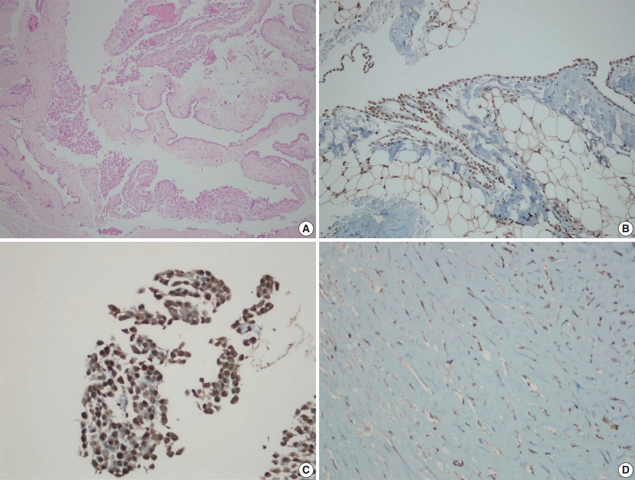
Malignant mesothelioma is a highly aggressive tumor that can be confused with a benign mesothelial lesion, especially cytomorphologic lesions. BRCA1-associated protein 1 (BAP1) acts as a tumor suppressor. In this study, we aim to investigate the value of BAP1 staining of malignant mesothelioma cases with expression loss and diagnosis in cell block and biopsy tissue.
Methods
Between January 2009 and March 2017, 64 mesotheliomas, 117 reactive mesothelial hyperplasias, and 20 fibrinous pleuritis/pericarditis were diagnosed with morphologic and immunohistochemical findings in our pathology clinic and were included in the study. Formalin-fixed, paraffin-embedded tissues were immunohistochemically examined for BAP1. Inflammatory and stromal cells were used as positive internal controls. BAP1 was assessed for nuclear staining in mesothelial cells.
Results
Examinations of the relationship between patient diagnosis and BAP1 biopsy status showed that the BAP1 loss rate (76.6%) was significantly higher in malignant mesothelioma cases than in other benign diseases (0%) (p<.001). Sensitivity and specificity were 76.56% and 100%, respectively, for biopsy tissue from malignant mesothelioma. Sensitivity and specificity were both 100% for BAP1 test on cell block tissue. Furthermore, the consistency between BAP1 cell block and biopsy results was excellent (ĸ=0.90) and the correlation was significant (p<.001).
Conclusions
This study shows that BAP1 expression loss in both cytology and biopsy tissue in biopsy-confirmed malignant mesothelioma cases is an essential parameter for malignant mesothelioma diagnosis. -
Citations
Citations to this article as recorded by
- Definitive diagnosis of pleural mesothelioma by pleural effusion cytology: the MesoCyto study
Masaki Hashimoto, Toshiyuki Minami, Ryuichi Waseda, Masaru Takenaka, Keigo Uchimura, Ayuko Sato, Takashi Kijima, Toshihiko Sato, Fumihiro Tanaka, Kazuhiro Yatera, Masaki Fujita, Masanori Hisaoka, Kazuki Nabeshima, Tohru Tsujimura, Seiki Hasegawa
International Journal of Lung Cancer.2026; 1(3): 100041. CrossRef - Diagnostic Challenges in the Pathological Approach to Pleural Mesothelioma
Stefano Lucà, Giovanna Pignata, Alessandro Cioce, Cecilia Salzillo, Rossella De Cecio, Gerardo Ferrara, Carminia Maria Della Corte, Floriana Morgillo, Alfonso Fiorelli, Marco Montella, Renato Franco
Cancers.2025; 17(3): 481. CrossRef - Thrombocytosis as a paraneoplastic syndrome in metastatic malignant peritoneal mesothelioma of biphasic morphology mimicking ovarian adenocarcinoma: A case report
Moustafa S. Alhamadh, Rakan B. Alanazi, Osama Mohaamad Wadaan, Abdulrahman Yousef Alhabeeb, Mohammad Alkaiyat, Ohoud Zaid Aljarbou, Fouad Sabatin
Clinical Case Reports.2023;[Epub] CrossRef - Primary cardiac mesothelioma presenting with fulminant recurrent pericarditis: a case report
Shmuel Schwartzenberg, Yaron Shapira, Victor Rubachevski, Ram Sharony, Harry Klimis, Domenico Filomena, Edgar Francisco Carrizales Sepulveda, Nikolaos Spinthakis, Jonathan Senior
European Heart Journal - Case Reports.2023;[Epub] CrossRef
- Definitive diagnosis of pleural mesothelioma by pleural effusion cytology: the MesoCyto study
- Foreign Body Reaction and Expression of Matrix Metalloproteinases/Tissue Inhibitor of Metalloproteinase by Injection of Mineral Fibers in Rats.
- Dong Kweon Seo, Jong Im Lee, Jung Ran Kim
- Korean J Pathol. 2011;45(6):604-611.
- DOI: https://doi.org/10.4132/KoreanJPathol.2011.45.6.604
- 4,031 View
- 25 Download
-
Abstract
PDF
- BACKGROUND
The host response to natural fibers results in granuloma formation in an effort to limit tissue destruction. Matrix metalloproteinases (MMPs) are important molecules in the inflammatory granulomatous or reparative reaction. Here, we studied the foreign body reaction that occurs following natural fibers implantation by investigating MMPs and tissue Inhibitor of MMPs (TIMPs) in an in vivo model.
METHODS
Female Sprague-Dawley rats were treated with crocidolite fiber or fibrous talc via subcutaneous and intraperitoneal injections and immunohistochemistry was conducted to confirm the expression of MMPs and TIMP-2 in tissue sections.
RESULTS
We identified that mineral fibers elicited granulomas. Fibrous talc or intraperitoneal injection resulted in larger granulomas and severe tissue destruction compared with the lesions induced by crocidolite or subcutaneous injection. The expression of MMPs was elevated while granulomatous lesions were formed. The relative levels of MMPs were lower in the talc injected or intraperitoneal route models than those of crocidolite injected or subcutaneous injection models during the entire experiment.
CONCLUSIONS
These findings demonstrate that specific expression of MMPs/TIMP is inversely related to the grade of tissue destruction and suggest that expression of MMPs is required for promoting granuloma formation and limiting tissue destruction.
Case Reports
- Analysis of Pulmonary Asbestos Body in Malignant Mesothelioma: A case report.
- Hoon Kyu Oh, Jae Yoe Ro, Chul Jong Yoon, Je Geun Chi
- Korean J Pathol. 1999;33(5):361-366.
- 2,316 View
- 20 Download
-
Abstract
PDF
- The association between occupational asbestos exposure and the subsequent development of malignant mesothelioma of pleura is well recognized. We analyzed an asbestos body by energy dispersive X-ray analyser in a case of malignant mesothelioma of pleura who had a history of asbestos exposure 30 years ago. In transmission electron microscope, the asbestos body was composed of a core of refractile thin asbestos fiber bundle and beaded masses of electron-dense iron and protein complex. The core fibers were analyzed as an amphibole type crocidolite fiber [(Na2Fe3Fe2(Si8O22)(OH)2] which composed of high content of silicon, iron and sodium.
- Rounded Atelectasis: A Brief Case Report.
- Gou Young Kim, Ji Young Park, Joung ho Han, Tae Seong Kim, Jhin gook Kim
- Korean J Pathol. 2003;37(4):279-281.
- 2,561 View
- 53 Download
-
Abstract
PDF
- Rounded atelectasis is a focal, pleural-based lesion that is the result of pleural and subpleural scarring and atelectasis of the adjacent lung tissue. We experienced a case of asbestosassociated rounded atelectasis that had developed in a 50-year-old male. When examined with routine chest radiography, the patient was shown to have an asymptomatic chest mass.Computed tomography showed a pleural-based mass with a curvilinear shape about 4.2 cmin greatest diameter in the medial basal segment of the right lower lobe. To exclude the possibilityof malignancy the mass was excised by video-assisted thoracotomy. The mass wasround and firm, and was gray and yellow in color. Microscopically, marked pleural fibrosisextended into the underlying lung parenchyme and then resulted in atelectasis. There areferruginous bodies in dense fibrous pleura.
 First
First Prev
Prev


