 E-submission
Search
- Page Path
- HOME > Search
Case Study
- Clinicopathological characteristics of digestive system angioleiomyomas: case report and literature review
- Georgios Kalliopitsas, Christos Topalidis, Constantine Halkias, Theodora Gkeka, Konstantinos Sapalidis, Triantafyllia Koletsa
- J Pathol Transl Med. 2025;59(6):453-459. Published online October 28, 2025
- DOI: https://doi.org/10.4132/jptm.2025.08.04
- 3,420 View
- 110 Download
-
 Abstract
 PDF
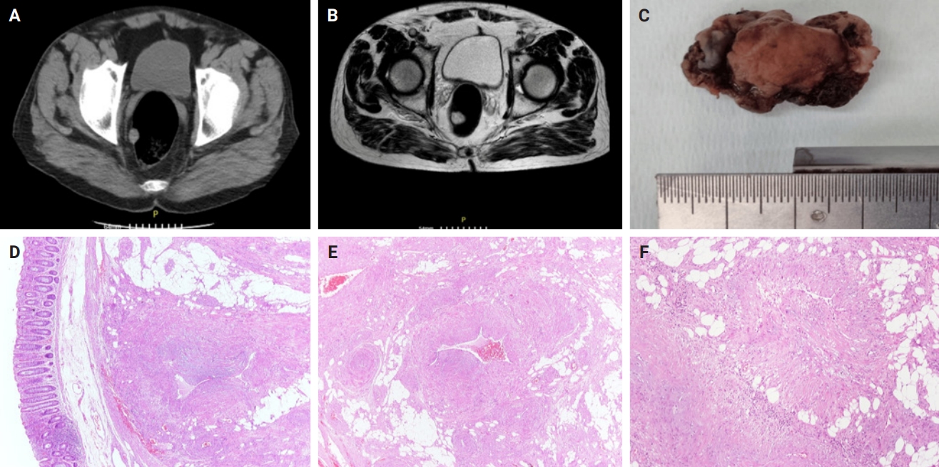
PDF - Angioleiomyomas are benign soft tissue tumors originating from the vascular wall. Although angioleiomyomas mainly occur in extremities, followed by head, neck, and trunk, they can also be found throughout the digestive system and especially in the oral cavity. Herein, the fourth case of a rectal angioleiomyoma in the English literature is reported and the clinicopathological features of digestive system angioleiomyomas were investigated. In contrast to their soft tissue counterparts, digestive system angioleiomyomas mainly affect males at a slightly younger age. Angioleiomyomas are mainly asymptomatic and only rarely elicit pain. Clinicians consider angioleiomyomas infrequently and instead include more common soft tissue or epithelial tumors in their differential diagnosis. To prevent angiomyolipoma misdiagnosis, pathologists should exercise caution when examining an angioleiomyoma composed of adipose tissue, smooth muscle, and blood vessels. Pathologists, radiologists, and surgeons should be aware that angioleiomyomas can occur in the digestive system.
Case Report
- Angiomyomatous Hamartoma of Popliteal Lymph Nodes Occurring in Association with Diffuse Pigmented Villonodular Synovitis of Knee.
- Hyun Soo Kim, Ki Yong Na, Jae Hoon Lee, Nam Su Cho, Gou Young Kim, Sung Jig Lim
- Korean J Pathol. 2011;45:S58-S61.
- DOI: https://doi.org/10.4132/KoreanJPathol.2011.45.S1.S58
- 4,018 View
- 20 Download
-
Abstract
PDF
- We report the first case of an angiomyomatous hamartoma (AH) of the popliteal lymph nodes (LNs) occurring in association with diffuse pigmented villonodular synovitis (PVNS) of the knee. AH is a rare benign vascular disease with a predisposition for the LNs of the inguinal region. Twenty-five cases of AH have been reported to date; however, the precise pathogenesis is still undetermined. In the present case, an open synovectomy revealed two of three popliteal LNs in close proximity to the extra-articular component of diffuse PVNS. These LNs demonstrated irregularly distributed thick-walled blood vessels in the hilum. These vessels extended into the medulla and cortex and were associated with haphazardly arranged smooth muscle cells in the sclerotic stroma. These findings are compatible with an AH. Our observations raise the possibility that AH of the popliteal LNs may represent an abnormal proliferative reaction against the inflammatory process caused by PVNS of the knee.
 First
First Prev
Prev


