 E-submission
Search
- Page Path
- HOME > Search
Original Articles
- A scoring system for the diagnosis of non-alcoholic steatohepatitis from liver biopsy
- Kyoungbun Lee, Eun Sun Jung, Eunsil Yu, Yun Kyung Kang, Mee-Yon Cho, Joon Mee Kim, Woo Sung Moon, Jin Sook Jeong, Cheol Keun Park, Jae-Bok Park, Dae Young Kang, Jin Hee Sohn, So-Young Jin
- J Pathol Transl Med. 2020;54(3):228-236. Published online April 15, 2020
- DOI: https://doi.org/10.4132/jptm.2020.03.07
- 15,940 View
- 283 Download
- 11 Web of Science
- 11 Crossref
-
 Abstract
 PDF
PDF - Background
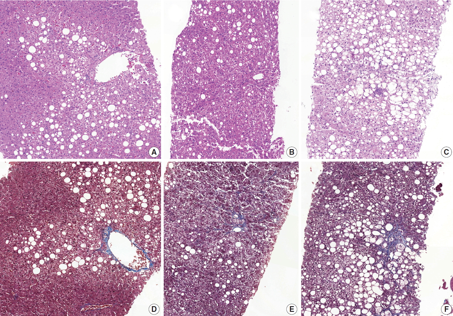
Liver biopsy is the essential method to diagnose non-alcoholic steatohepatitis (NASH), but histological features of NASH are too subjective to achieve reproducible diagnoses in early stages of disease. We aimed to identify the key histological features of NASH and devise a scoring model for diagnosis.
Methods
Thirteen pathologists blindly assessed 12 histological factors and final histological diagnoses (‘not-NASH,’ ‘borderline,’ and ‘NASH’) of 31 liver biopsies that were diagnosed as non-alcoholic fatty liver disease (NAFLD) or NASH before and after consensus. The main histological parameters to diagnose NASH were selected based on histological diagnoses and the diagnostic accuracy and agreement of 12 scoring models were compared for final diagnosis and the NAFLD Activity Score (NAS) system.
Results
Inter-observer agreement of final diagnosis was fair (κ = 0.25) before consensus and slightly improved after consensus (κ = 0.33). Steatosis at more than 5% was the essential parameter for diagnosis. Major diagnostic factors for diagnosis were fibrosis except 1C grade and presence of ballooned cells. Minor diagnostic factors were lobular inflammation ( ≥ 2 foci/ × 200 field), microgranuloma, and glycogenated nuclei. All 12 models showed higher inter-observer agreement rates than NAS and post-consensus diagnosis (κ = 0.52–0.69 vs. 0.33). Considering the reproducibility of factors and practicability of the model, summation of the scores of major (× 2) and minor factors may be used for the practical diagnosis of NASH.
Conclusions
A scoring system for the diagnosis of NAFLD would be helpful as guidelines for pathologists and clinicians by improving the reproducibility of histological diagnosis of NAFLD. -
Citations
Citations to this article as recorded by
- Preclinical liver toxicity models: Advantages, limitations and recommendations
Devaraj Ezhilarasan, Sivanesan Karthikeyan, Mustapha Najimi, Paramasivan Vijayalakshmi, Ganapathy Bhavani, Muthukrishnan Jansi Rani
Toxicology.2025; 511: 154020. CrossRef - Hepatic miR-93 promotes the pathogenesis of metabolic dysfunction-associated steatotic liver disease by suppressing SIRT1
Yo Han Lee, Jinyoung Lee, Joonho Jeong, Kieun Park, Bukyung Baik, Yuseong Kwon, Kimyeong Kim, Keon Woo Khim, Haneul Ji, Ji Young Lee, Kwangho Kim, Ji Won Kim, Tam Dao, Misung Kim, Tae Young Lee, Yong Ryoul Yang, Haejin Yoon, Dongryeol Ryu, Seonghwan Hwang
Metabolism.2025; 169: 156266. CrossRef - Deep learning-based method for grading histopathological liver fibrosis in rodent models of metabolic dysfunction-associated steatohepatitis
Soo Min Ko, Jae-ik Shin, Yiyu Hong, Hyunji Kim, Insuk Sohn, Ji-Young Lee, Hyo-Jeong Han, Da Som Jeong, Yerin Lee, Woo-Chan Son
Frontiers in Medicine.2025;[Epub] CrossRef - Liver biopsy in the modern era: from traditional techniques to artificial intelligence and multi-omics integration
Nasar Alwahaibi, Maryam Alwahaibi
Frontiers in Medicine.2025;[Epub] CrossRef - Presurgery health influences outcomes following vertical sleeve gastrectomy in adolescents
Debi Swertfeger, Ahlee Kim, Hannah Sexmith, Maria E. Moreno‐Fernandez, W. Sean Davidson, Michael Helmrath, Todd Jenkins, Tsuyoshi Okura, Esmond Geh, Stavra A. Xanthakos, Sara Szabo, Takahisa Nakamura, Senad Divanovic, Amy Sanghavi Shah
Obesity.2024; 32(6): 1187. CrossRef - Immunobiotic Bacteria Attenuate Hepatic Fibrosis through the Modulation of Gut Microbiota and the Activation of Aryl‐Hydrocarbon Receptors Pathway in Non‐Alcoholic Steatohepatitis Mice
Paulraj Kanmani, Julio Villena, Soo‐kyoung Lim, Eun‐Ji Song, Young‐Do Nam, Hojun Kim
Molecular Nutrition & Food Research.2024;[Epub] CrossRef - Lipid nanoparticle-mediated hepatocyte delivery of siRNA and silibinin in metabolic dysfunction-associated steatotic liver disease
Yifu Lyu, Xiuyi Yang, Lei Yang, Jinyu Dai, Huanyu Qin, Yunuo Zhou, Yunan Huang, Yanmei Wang, Di Wu, Qindai Shuai, Qilong Li, Xiaofei Xin, Lifang Yin
Journal of Controlled Release.2024; 373: 385. CrossRef - Enhanced hepatoprotective effects of empagliflozin and vitamin D dual therapy against metabolic dysfunction‐associated steatohepatitis in mice by boosted modulation of metabolic, oxidative stress, and inflammatory pathways
Wesam F. Farrash, Shakir Idris, Mohamed E. Elzubier, Elshiekh B. A. Khidir, Akhmed Aslam, Abdulrahman Mujalli, Riyad A. Almaimani, Ahmad A. Obaid, Mahmoud Z. El‐Readi, Mohammad A. Alobaidy, Afnan Salaka, Afnan M. Shakoori, Alaa M. Saleh, Faisal Minshawi,
International Journal of Experimental Pathology.2024; 105(6): 219. CrossRef - Bilirubin, a hepatoprotective agent that activates SIRT1, PGC-1α, and PPAR-α, while inhibiting NF-κB in rats with metabolic-associated fatty liver disease
Motahareh Taghizadeh, Mohammad Hasan Maleki, Omid Vakili, Ramin Tavakoli, Parvin Zarei, Amirreza Dehghanian, Hossein Bordbar, Sayed Mohammad Shafiee
Scientific Reports.2024;[Epub] CrossRef - Changes in indications for outpatient percutaneous liver biopsy over 5 years: from hepatitis C to fatty liver disease
Marlone Cunha-Silva, Luíza D. Torres, Mariana F. Fernandes, Tirzah de M. Lopes Secundo, Marina C.G. Moreira, Ademar Yamanaka, Leonardo T. Monici, Larissa B. Eloy da Costa, Daniel F. Mazo, Tiago Sevá-Pereira
Gastroenterología y Hepatología.2022; 45(8): 579. CrossRef - Changes in indications for outpatient percutaneous liver biopsy over 5 years: from hepatitis C to fatty liver disease
Marlone Cunha-Silva, Luíza D. Torres, Mariana F. Fernandes, Tirzah de M. Lopes Secundo, Marina C.G. Moreira, Ademar Yamanaka, Leonardo T. Monici, Larissa B. Eloy da Costa, Daniel F. Mazo, Tiago Sevá-Pereira
Gastroenterología y Hepatología (English Edition).2022; 45(8): 579. CrossRef
- Preclinical liver toxicity models: Advantages, limitations and recommendations
- Interobserver Agreement on Pathologic Features of Liver Biopsy Tissue in Patients with Nonalcoholic Fatty Liver Disease
- Eun Sun Jung, Kyoungbun Lee, Eunsil Yu, Yun Kyung Kang, Mee-Yon Cho, Joon Mee Kim, Woo Sung Moon, Jin Sook Jeong, Cheol Keun Park, Jae-Bok Park, Dae Young Kang, Jin Hee Sohn, So-Young Jin
- J Pathol Transl Med. 2016;50(3):190-196. Published online April 18, 2016
- DOI: https://doi.org/10.4132/jptm.2016.03.01
- 15,557 View
- 276 Download
- 29 Web of Science
- 30 Crossref
-
Abstract
PDF
- Background
The histomorphologic criteria for the pathological features of liver tissue from patients with non-alcoholic fatty liver disease (NAFLD) remain subjective, causing confusion among pathologists and clinicians. In this report, we studied interobserver agreement of NAFLD pathologic features and analyzed causes of disagreement.
Methods
Thirty-one cases of clinicopathologically diagnosed NAFLD from 10 hospitals were selected. One hematoxylin and eosin and one Masson’s trichrome-stained virtual slide from each case were blindly reviewed with regard to 12 histological parameters by 13 pathologists in a gastrointestinal study group of the Korean Society of Pathologists. After the first review, we analyzed the causes of disagreement and defined detailed morphological criteria. The glass slides from each case were reviewed a second time after a consensus meeting. The degree of interobserver agreement was determined by multi-rater kappa statistics.
Results
Kappa values of the first review ranged from 0.0091–0.7618. Acidophilic bodies (k = 0.7618) and portal inflammation (k = 0.5914) showed high levels of agreement, whereas microgranuloma (k = 0.0984) and microvesicular fatty change (k = 0.0091) showed low levels of agreement. After the second review, the kappa values of the four major pathological features increased from 0.3830 to 0.5638 for steatosis grade, from 0.1398 to 0.2815 for lobular inflammation, from 0.1923 to 0.3362 for ballooning degeneration, and from 0.3303 to 0.4664 for fibrosis.
Conclusions
More detailed histomorphological criteria must be defined for correct diagnosis and high interobserver agreement of NAFLD. -
Citations
Citations to this article as recorded by- Recent Advances in the Application of Machine Learning Models in Metabolic Dysfunction–Associated Steatotic Liver Disease
Fang Yang, Xueyue Sun, Kui Jiang, Mingxin Zhang, Chao Sun
Diabetes/Metabolism Research and Reviews.2026;[Epub] CrossRef - Double Graph Attention Network for predicting non-alcoholic fatty liver disease in patients with type 2 diabetes
Tianbin Chen, Yongbin Zeng, Jinlin Wang, Xiao Sun, Sihao Liu, Ya Fu, Qiang Yi, Qishui Ou, Kai Yan, Zhiheng Zhou
Artificial Intelligence in Medicine.2026; 174: 103369. CrossRef - Liver Biopsy in Metabolic-Associated Steatotic Liver Disease: Accuracy, Challenges, and Alternatives
Luca Borz-Baba, Adem Aydin, Russell Parvin
Cureus.2026;[Epub] CrossRef - Quantitative regression of qFibrosis with resmetirom: Exploratory histologic endpoints from the MAESTRO-NASH phase III clinical trial
Jörn M. Schattenberg, Pierre Bedossa, Cynthia D. Guy, Rebecca Taub, Dominic Labriola, Hang Zhang, James Hennan, Raul C. Camacho, Elaine Chng, Yayun Ren, Dean Tai, Stephen A. Harrison
Journal of Hepatology.2026;[Epub] CrossRef - Chronic polypharmacy, monotherapy, and deprescribing: Understanding complex effects on the hepatic proteome of aging mice
Kevin Winardi, John Mach, Matthew J. McKay, Mark P. Molloy, Sarah J. Mitchell, Michael R. MacArthur, Catriona McKenzie, David G. Le Couteur, Sarah N. Hilmer
Aging Cell.2025;[Epub] CrossRef - Utility of AI digital pathology as an aid for pathologists scoring fibrosis in MASH
Desiree Abdurrachim, Serene Lek, Charlene Zhi Lin Ong, Chun Kit Wong, Yongqi Zhou, Aileen Wee, Gwyneth Soon, Timothy J. Kendall, Michael O. Idowu, Christopher Hendra, Ashmita Saigal, Radha Krishnan, Elaine Chng, Dean Tai, Gideon Ho, Thomas Forest, Annaswa
Journal of Hepatology.2025; 82(5): 898. CrossRef - Artificial intelligence scoring of liver biopsies in a phase II trial of semaglutide in nonalcoholic steatohepatitis
Vlad Ratziu, Sven Francque, Cynthia A. Behling, Vanja Cejvanovic, Helena Cortez-Pinto, Janani S. Iyer, Niels Krarup, Quang Le, Anne-Sophie Sejling, Dina Tiniakos, Stephen A. Harrison
Hepatology.2024; 80(1): 173. CrossRef - Classification of the Stages of Nonalcoholic Steatohepatitis via Federated General Visual Representation Learning
Mehmet Nergiz
International Journal of Imaging Systems and Technology.2024;[Epub] CrossRef - Outcome prediction in metabolic dysfunction‐associated steatotic liver disease using stain‐free digital pathological assessment
Timothy J. Kendall, Elaine Chng, Yayun Ren, Dean Tai, Gideon Ho, Jonathan A. Fallowfield
Liver International.2024; 44(10): 2511. CrossRef - Non-alcoholic fatty liver disease: the pathologist’s perspective
Wei-Qiang Leow, Anthony Wing-Hung Chan, Paulo Giovanni L. Mendoza, Regina Lo, Kihan Yap, Haeryoung Kim
Clinical and Molecular Hepatology.2023; 29(Suppl): S302. CrossRef - CT-based Hounsfield unit values reflect the degree of steatohepatitis in patients with low-grade fatty liver disease
Ha Neul Kim, Hong Jae Jeon, Hei Gwon Choi, In Sun Kwon, Woo Sun Rou, Jeong Eun Lee, Tae Hee Lee, Seok Hyun Kim, Byung Seok Lee, Kyung Sook Shin, Hyun Jung Lee, Hyuk Soo Eun
BMC Gastroenterology.2023;[Epub] CrossRef - Artificial intelligence and deep learning: New tools for histopathological diagnosis of nonalcoholic fatty liver disease/nonalcoholic steatohepatitis
Yoshihisa Takahashi, Erdenetsogt Dungubat, Hiroyuki Kusano, Toshio Fukusato
Computational and Structural Biotechnology Journal.2023; 21: 2495. CrossRef - An integrated gene-to-outcome multimodal database for metabolic dysfunction-associated steatotic liver disease
Timothy J. Kendall, Maria Jimenez-Ramos, Frances Turner, Prakash Ramachandran, Jessica Minnier, Michael D. McColgan, Masood Alam, Harriet Ellis, Donald R. Dunbar, Gabriele Kohnen, Prakash Konanahalli, Karin A. Oien, Lucia Bandiera, Filippo Menolascina, An
Nature Medicine.2023; 29(11): 2939. CrossRef - Improved pathology reporting in NAFLD/NASH for clinical trials
Caitlin Rose Langford, Marc H Goldinger, Darren Treanor, Clare McGenity, Jonathan R Dillman, Daniela S Allende, Robert Goldin, Elizabeth M Brunt, Kurt Zatloukal, Helmut Denk, Kenneth A Fleming
Journal of Clinical Pathology.2022; 75(2): 73. CrossRef - Standardizing the histological assessment of late posttransplantation biopsies from pediatric liver allograft recipients
Stefan G. Hübscher, Sandy Feng, Annette S. H. Gouw, Hironori Haga, Hyo Jeong Kang, Deirdre A. Kelly, Mina Komuta, Andrew Lesniak, Benjamin A. Popp, Henkjan J. Verkade, Eunsil Yu, Anthony J. Demetris
Liver Transplantation.2022; 28(9): 1475. CrossRef - Discordant pathological diagnosis of non‐alcoholic fatty liver disease: A prospective multicenter study
Takuya Kuwashiro, Hirokazu Takahashi, Hideyuki Hyogo, Yuji Ogawa, Kento Imajo, Masato Yoneda, Takashi Nakahara, Satoshi Oeda, Kenichi Tanaka, Yuichiro Amano, Shinji Ogawa, Atsushi Kawaguchi, Shinichi Aishima, Masayoshi Kage, Kazuaki Chayama, Atsushi Nakaj
JGH Open.2020; 4(3): 497. CrossRef - Obeticholic acid for the treatment of nonalcoholic steatohepatitis: Expectations and concerns
Stergios A. Polyzos, Jannis Kountouras, Christos S. Mantzoros
Metabolism.2020; 104: 154144. CrossRef - A scoring system for the diagnosis of non-alcoholic steatohepatitis from liver biopsy
Kyoungbun Lee, Eun Sun Jung, Eunsil Yu, Yun Kyung Kang, Mee-Yon Cho, Joon Mee Kim, Woo Sung Moon, Jin Sook Jeong, Cheol Keun Park, Jae-Bok Park, Dae Young Kang, Jin Hee Sohn, So-Young Jin
Journal of Pathology and Translational Medicine.2020; 54(3): 228. CrossRef - An Improved qFibrosis Algorithm for Precise Screening and Enrollment into Non-Alcoholic Steatohepatitis (NASH) Clinical Trials
Wei-Qiang Leow, Pierre Bedossa, Feng Liu, Lai Wei, Kiat-Hon Lim, Wei-Keat Wan, Yayun Ren, Jason Pik-Eu Chang, Chee-Kiat Tan, Aileen Wee, George Boon-Bee Goh
Diagnostics.2020; 10(9): 643. CrossRef - Deep learning quantification of percent steatosis in donor liver biopsy frozen sections
Lulu Sun, Jon N. Marsh, Matthew K. Matlock, Ling Chen, Joseph P. Gaut, Elizabeth M. Brunt, S. Joshua Swamidass, Ta-Chiang Liu
EBioMedicine.2020; 60: 103029. CrossRef - Magnetic resonance elastography SE-EPI vs GRE sequences at 3T in a pediatric population with liver disease
Juan S. Calle-Toro, Suraj D. Serai, Erum A. Hartung, David J. Goldberg, Bradley D. Bolster, Kassa Darge, Sudha A. Anupindi
Abdominal Radiology.2019; 44(3): 894. CrossRef - R2 relaxometry based MR imaging for estimation of liver iron content: A comparison between two methods
Juan S. Calle-Toro, Christian A. Barrera, Dmitry Khrichenko, Hansel J. Otero, Suraj D. Serai
Abdominal Radiology.2019; 44(9): 3058. CrossRef - Inhibition of mitochondrial fatty acid oxidation in drug-induced hepatic steatosis
Bernard Fromenty
Liver Research.2019; 3(3-4): 157. CrossRef - Standardising the interpretation of liver biopsies in non‐alcoholic fatty liver disease clinical trials
Rish K. Pai, David E. Kleiner, John Hart, Oyedele A. Adeyi, Andrew D. Clouston, Cynthia A. Behling, Dhanpat Jain, Sanjay Kakar, Mayur Brahmania, Lawrence Burgart, Kenneth P. Batts, Mark A. Valasek, Michael S. Torbenson, Maha Guindi, Hanlin L. Wang, Veeral
Alimentary Pharmacology & Therapeutics.2019; 50(10): 1100. CrossRef - NAFLD Histology: a Critical Review and Comparison of Scoring Systems
Rish K. Pai
Current Hepatology Reports.2019; 18(4): 473. CrossRef - Hepatic sonic hedgehog protein expression measured by computer assisted morphometry significantly correlates with features of non-alcoholic steatohepatitis
Michael Estep, Rohini Mehta, Gary Bratthauer, Lakshmi Alaparthi, Fanny Monge, Simon Ali, Dinan Abdelatif, Zahra Younoszai, Maria Stepanova, Zachary D. Goodman, Zobair M. Younossi
BMC Gastroenterology.2019;[Epub] CrossRef - Validation of intimate correlation between visceral fat and hepatic steatosis: Quantitative measurement techniques using CT for area of fat and MR for hepatic steatosis
Moon Hyung Choi, Joon-Il Choi, Michael Yong Park, Sung Eun Rha, Soon Nam Oh, Seung Eun Jung, Jae Young Byun, Stephan Kannengiesser, Yohan Son
Clinical Nutrition.2018; 37(1): 214. CrossRef - Ultrasound or MR elastography of liver: which one shall I use?
Meng Yin, Sudhakar K. Venkatesh
Abdominal Radiology.2018; 43(7): 1546. CrossRef - Feasibility and agreement of stiffness measurements using gradient-echo and spin-echo MR elastography sequences in unselected patients undergoing liver MRI
Guilherme Moura Cunha, Kevin J Glaser, Anke Bergman, Rodrigo P Luz, Eduardo H de Figueiredo, Flavia Paiva Proença Lobo Lopes
The British Journal of Radiology.2018; : 20180126. CrossRef - Second harmonic generation microscopy provides accurate automated staging of liver fibrosis in patients with non-alcoholic fatty liver disease
Pik Eu Chang, George Boon Bee Goh, Wei Qiang Leow, Liang Shen, Kiat Hon Lim, Chee Kiat Tan, Manlio Vinciguerra
PLOS ONE.2018; 13(6): e0199166. CrossRef
- Recent Advances in the Application of Machine Learning Models in Metabolic Dysfunction–Associated Steatotic Liver Disease
- Histologic Pattern of Alcoholic Liver Disease in Korea.
- Chan Il Park, Ho Guen Kim, So Young Jin, Mi Kyung Lee, Yoo Bock Lee
- Korean J Pathol. 1989;23(3):292-304.
- 2,312 View
- 21 Download
-
Abstract
PDF
- To elucidate the histologic pattern of alcoholic liver disease (ALD) in Korea, liver biopsies from 173 chronic alcoholics with clinical liver diseases were classified according to the pathologic parameters. One hundred and seventeen cases, the sum of 91 of 116 serum HBsAg negative and 26 of 57 HBsAg positive patients, had the histologic evidence of ALD. Fatty change(23.9%), alcoholic fibrosis (AF)(23.1%) and cirrhosis (23.1%), comprised the three major ALDs, and only 8.5% of cases fit the criteria of alcoholic hepatitis. Chronic sclerosing hyaline disease (CSHD), chronic active alcoholic hepatitis (CAAH) and AF, where non-cirrhotic fibrosis is the predominant change, comprised 44.5% of ALD. Both features of ALD and HBV liver disease (HBV-LD) were found in 17 cases that included 8 AF and 7 cirrhosis. These 17 patients tended to consume less alcohol than patients with other types of pure ALD except alcoholic heaptitis. Patients with the serum HBsAg positive ALD (37.4years) were about 8 years younger than those with the serum HBsAg negative ALD (45.1years). More or less fatty change and foamy degeneration were seen in 77.4% and 31.6% of ALD respectively. Mallory bodies, megamitochondria, iron deposition and perihepatocellular fibrosis were found in 20.5%, 29.9%, 42.7% and 77.8%, respectively. These findings indicate that non-cirrhotic chronic ALD such as CSHD, CAAH and AF are the important histologic patterns of ALD in Korea, and that chronic alcohol consumption and HBV may act synergistically in developing liver disease.
- Alcoholic Type Cirrhosis Following Side to Side Ileo-Transverse Colon Anastomosis.
- Kwang Hwa Park, Kwang Hyup Han, Chan Il Park
- Korean J Pathol. 1990;24(2):148-152.
- 2,129 View
- 13 Download
-
Abstract
PDF
- A case of micronodular cirrhosis of the alcoholic type developed following an intestianl bypass surgery in a 47 year-old nonalcoholic male patient is presented. The patient denied any drug use of a long duration and had no diabetes mellitus. Five years before, a side to side ileo-transverse colon anastomosis had been performed for perforated intestinal tuberculosis at 1 m proximal to the ileocecal valve, bypassing a short segment of ileum (about 1.5 m) and transverse colon. The ileum distal to the perforated site had been found completely stenosed. He was severely lean with evidences of nutritional deficiency such as low serum levels of the albumin and vitamin B12. The liver biopsy showed a fatty change, Mallory bodies and perihepatocellular collagenosis within the cirrhotic nodules. The present case suggests that, when there are blind loop formation and nutritional deficiency, hepatic changes identical to those following jejunoileal bypass could develop even by reduction of a shore segment of the small intestine.
- Ultrastructural Study of Alcohol-Induced Gastric Mucosal Change of Rat.
- Kam Rae Cho, Kun Young Kwon, Eun Sook Chang
- Korean J Pathol. 1993;27(4):362-370.
- 2,328 View
- 25 Download
-
Abstract
PDF
- In an attempt ultrastructural study of alcohol-induced gastric mucosal change, we selected sixty Sprague-Dawley rats. The rats were administrated with 4 ml of 10% and 40% ethanol enterically and examined by light and electron microscopy. Light microscopically, the thickness of the mucus layer of both 10% and 40% ethanol groups was decreased. The antral mucosa revealed focal inflammatory infiltrates, disturbed glandular arrangements, and significant decrease of mucosal thichness and proper glands. On scanning electron microscopy, flattened or swollen mucosal epithelium and irregularly distributed gastric pits were seen in both experimental groups, and these changes were more severe in the groups of higher concentration and longer duration. On transmission electron microscopy, mitochondrial abnormalities with myelin-like materials and dilatation of endoplasmic reticulum and cytoplasmic blebs were observed. Also the mucus cells show significantly decreased mucus globules, increased fat vacuoles, and large autophagic vacuoles. These alterations were similar to those produced by ethanol in the liver and small intestine. This study indicates that, prolonged administration of ethanol induced chronic gastritis, especially chronic atrophic gastritis.
- Liver Cirrhosis: Etiological diagnosis and morphological characteristics of 369 biopsy-proven cases.
- Eun Kyung Han, Chanil Park, Sang In Lee
- Korean J Pathol. 1990;24(4):412-422.
- 2,942 View
- 37 Download
-
Abstract
PDF
- To pursue a desirable format for the pathological diagnosis of liver cirrhosis, the authors attempted to classify 369 biopsy-proven cirrhosis on the basis of etiology and made effort to find out the morphological characteristics of each category. About 735 of total cases were HBsAg seropositive postnecrotic cirrhosis. Alcholic cirrhosis ws the second most frequent type, although accounted only 6.8%. In about 15%, the etiology was not known. Excluding the congenital biliary atresia, chronic biliary obstruction appeared to be a rare cause of cirrhosis among these biopsied cases. Of the HBsAg positive postnecrotic cirrhosis, the eAg seropositive cases tended to be micronodular and to show a higher necroinflammatory activity, in contrast to eAg seronegative cases and those complicated by hepatocellular carcinoma (HCC), suggesting that the loss of eAg is followed by a decrease of the destructive activity, active regeneration of hepatocytes and finally the development of HCC. alcoholic cirrhosis was micronodular in 64% and revealed histologic evidences of alcoholic liver disease in most cases. The results indicate that etiological diagnosis can be made in most cases of cirrhosis by the morphological characteristics and the precise clinical informations, including those on the NANB virus and the inborn error of metabolism, and that the pathological diagnosis should be more comprehensive, implicating the etiology, the nodular size and the necroinflammatory activity.
- A Clinicopathologic Study on Chronic Alcoholic Hepatitis.
- Gi Yeong Huh, Sun Kyung Lee
- Korean J Pathol. 1988;22(4):393-403.
- 2,024 View
- 14 Download
-
Abstract
PDF
- This study was undertaken to evaluate the significant diagnostic points of chronic alcoholic hepatitis (CALH) among clinicopathologic findings observed. The specimens used in this study were 20 cases of CALH and 28 cases of chronic active viral hepatitis (CAVH), which were diagnosed at our University Hospital during 9 years period from 1978 to 1987. In these cases, comparative analysis of age and sex distribution, major clinical manifestations, and laboratory and histopathologic findings was performed. The results obained were summarized as follows: Among 20 cases of CALH, the sex distribution was 15 in male and 15 in female with a ratio of 3:1. The range of age distribution was wide from third to seventh decade. There was no recognizable special point about the age and sex distribution of CALH, compared with cases of CAVH. Major clinical manifestations of CALH were hepatomegaly (85%), jaundice (75%) and abdominal pain (50%). Also there was no recognizable special point about the major clinical manifestations of CALH, compared with cases of CAVH. Abnormal values of major laboratory items in CALH were observed in activities of serum r-GTP (100%), SGOT (95%), SGPT (75%) and serum alkaline phosphatase (60%), and total serum bilirubin (60%). Compared with CAVH in major laboratory findings, the significant diagnostic items of CALH were the activities of serum r-GTP and alkaline phosphatase. The characteristic histopathologic findings of CALH, which were compared with CAVH and observed in liver parenchyma, were fatty change (100%), cytoplasmic ballooning and coagulation (100%), delicate fibrosis (85%), bile stagnation (35%), and Mallory bodies (20%), and that observed blurring of limiting plate (60%) in portal and periportal areas.
 First
First Prev
Prev


