 E-submission
Search
- Page Path
- HOME > Search
Original Articles
- Spectrum of thyroiditis types: clinical, cytomorphological, and radiological findings
- Anam Singh, Indrajeet Kundu
- J Pathol Transl Med. 2025;59(6):421-433. Published online November 6, 2025
- DOI: https://doi.org/10.4132/jptm.2025.08.13

- 5,361 View
- 223 Download
-
 Abstract
 PDF
PDF - Background
Thyroiditis encompasses a range of inflammatory conditions affecting the thyroid gland. Lymphocytic thyroiditis (LT) is a common form of thyroiditis, with acute suppuration of the thyroid, while tuberculous thyroiditis is relatively rare. Fine-needle aspiration cytology (FNAC) remains a safe and cost-effective tool for diagnosing thyroid-related diseases, especially when paired with ultrasound (US) and clinical examination. Methods: This is a cross-sectional study including 21 cases. The cases were reported as thyroiditis on US and FNAC, and the findings were correlated with patient clinical history, symptoms during presentation, and serological profiles. Results: The cases of thyroiditis encompassed the more common forms, LT and subacute granulomatous thyroiditis (SAT), as well as relatively rare forms like tuberculous thyroiditis and thyroid abscess. Cases of follicular neoplasms (FN) arising in the context of LT also are included in this study. The case of tuberculous thyroiditis presented as a bulky thyroid gland that appeared heterogeneous on US with extensive necrosis on FNAC. The cases of thyroid abscess and SAT presented with painful neck swellings, with granulomas in the latter cases. US features of LT showed an array of appearances ranging from pseudonodular to an atrophic thyroid gland. All cases of FN showed a lymphocytic background. Conclusions: Thyroiditis is a commonly encountered condition that needs to be sub-categorized accurately into acute, subacute, and chronic types for appropriate clinical management, as they can sometimes show overlapping features. Though rare, acute suppurative and tuberculous thyroiditis are often encountered and warrant immediate care and treatment.
- Blocking Toll-like receptor 9 attenuates bleomycin-induced pulmonary injury
- Badr Alzahrani, Mohamed M. S. Gaballa, Ahmed A. Tantawy, Maha A. Moussa, Salma A. Shoulah, Said M. Elshafae
- J Pathol Transl Med. 2022;56(2):81-91. Published online March 2, 2022
- DOI: https://doi.org/10.4132/jptm.2021.12.27

- 9,357 View
- 157 Download
- 13 Web of Science
- 13 Crossref
-
Abstract
PDF
- Background
Acute respiratory distress syndrome (ARDS) is one of the most common complications in coronavirus disease 2019 patients suffering from acute lung injury (ALI). In ARDS, marked distortion of pulmonary architecture has been reported. The pulmonary lesions in ARDS include hemodynamic derangements (such as alveolar edema and hemorrhage), vascular and bronchiolar damage, interstitial inflammatory cellular aggregations, and eventually fibrosis. Bleomycin induces ARDS-representative pulmonary damage in mice and rats; therefore, we used bleomycin model mice in our study. Recently, Toll-like receptor 9 (TLR9) was implicated in the development of ARDS and ALI.
Methods
In this study, we evaluated the efficiency of a TLR9 blocker (ODN2088) on bleomycin-induced pulmonary damage. We measured the apoptosis rate, inflammatory reaction, and fibroplasia in bleomycin- and bleomycin + ODN2088-treated mice.
Results
Our results showed a significant amelioration in bleomycin-induced damage to pulmonary architecture following ODN2088 treatment. A marked decrease in pulmonary epithelial and endothelial apoptosis rate as measured by cleaved caspase-3 expression, inflammatory reaction as indicated by tumor necrosis factor α expression, and pulmonary fibrosis as demonstrated by Van Gieson staining and α-smooth muscle actin immunohistochemistry were observed following ODN2088 treatment.
Conclusions
All these findings indicate that blocking downstream TLR9 signaling could be beneficial in prevention or mitigation of ARDS through hemodynamic derangements, inflammation, apoptosis, and fibrosis. -
Citations
Citations to this article as recorded by
- Nano-sized DNase scavenges cell-free DNA for acute lung injury treatment
Ruijie Chen, Yitianhe Xu, Zhanzheng Ye, Yixuan Zhu, Mengxue Zhang, Fangfang Lv, Yunzhi Wang, Xinyu Di, Yinhao Lin, Shengnan Song, Zihao Huang, Shize Li, Zhinan He, Hailin Zhang, Longfa Kou
Journal of Controlled Release.2026; 394: 114846. CrossRef - Pharmacological repurposing of cilostazol to attenuate the progression of pulmonary fibrosis: efficacy validation via integrated network pharmacology and in vivo experimentation
Pranaya L. Misar, Kishor V. Otari, Vishal V. Pande, Pradyumna P. Ige
Naunyn-Schmiedeberg's Archives of Pharmacology.2026;[Epub] CrossRef - A novel mouse model of myositis-associated interstitial lung disease was established by using TLR9 agonist combined with muscle homogenate
Ling Bai, Jiarui Zhu, Wenlan Ma, Peipei Zhao, Feifei Li, Cen Zhang, Sigong Zhang
Clinical and Experimental Immunology.2025;[Epub] CrossRef - Toll-like Receptor 9 Inhibition Mitigates Fibroproliferative Responses in Translational Models of Pulmonary Fibrosis
Glenda Trujillo, Alicia Regueiro-Ren, Chunjian Liu, Buqu Hu, Ying Sun, Farida Ahangari, Vitoria Fiorini, Genta Ishikawa, Karam Al Jumaily, Johad Khoury, John McGovern, Chris J. Lee, Xue Yan Peng, Taylor Pivarnik, Huanxing Sun, Anjali Walia, Samuel Woo, Sh
American Journal of Respiratory and Critical Care Medicine.2025; 211(1): 91. CrossRef - CD103+ dendritic cell–fibroblast crosstalk via TLR9, TDO2, and AHR signaling drives lung fibrogenesis
Hannah Carter, Rita Medina Costa, Taylor S. Adams, Talon M. Gilchrist, Claire E. Emch, Monica Bame, Justin M. Oldham, Steven K. Huang, Angela L. Linderholm, Imre Noth, Naftali Kaminski, Bethany B. Moore, Stephen J. Gurczynski
JCI Insight.2025;[Epub] CrossRef - The Mitigated Effect of the Combination of Metformin and Stearic Acid to Ameliorate Bleomycin‐Induced Pulmonary Fibrosis in Rats via Inhibiting Gal‐3/Smad3/α‐SMA and TNF‐α/NF‐κβ Signaling Pathways
Maha M. Salem, Nermin S. Youssef, Mai El Keiy, Abeer A. Khamis
Journal of Biochemical and Molecular Toxicology.2025;[Epub] CrossRef - Unraveling the Interactive Role of Mitochondrial Dysfunction in Promoting Macrophage Polarization in Pulmonary Fibrosis
Jiajia Zou, Junling Jian, Dehua Ge, Bin Zhou, Jiaxiang Zhang
Journal of Biochemical and Molecular Toxicology.2025;[Epub] CrossRef - Mechanisms underlying dose-limiting toxicities of conventional chemotherapeutic agents
Mohammad Amin Manavi, Mohammad Hosein Fathian Nasab, Razieh Mohammad Jafari, Ahmad Reza Dehpour
Journal of Chemotherapy.2024; 36(8): 623. CrossRef - Innate Immune Response-Mediated Inflammation in Viral Pneumonia
Weiwei Ni, Xin Wei, Rui Wu
Journal of Pediatric Infectious Diseases.2024; 19(03): 140. CrossRef - Combination of losartan with pirfenidone: a protective anti-fibrotic against pulmonary fibrosis induced by bleomycin in rats
Arian Amirkhosravi, Maryamossadat Mirtajaddini Goki, Mahmoud Reza Heidari, Somayyeh Karami-Mohajeri, Maryam Iranpour, Maryam Torshabi, Mitra Mehrabani, Ali Mandegary, Mehrnaz Mehrabani
Scientific Reports.2024;[Epub] CrossRef - Suppression of miR-17 Alleviates Acute Respiratory Distress-associated Lung Fibrosis by Regulating Mfn2
Mei-xia Xu, Tao Xu, Ning An
Current Medical Science.2024; 44(5): 964. CrossRef - Study of Recombinant Interleukin-1 Receptor Antagonist Compositions Biological Activity After Injection and Inhalation in Mouse Model of Pulmonary Inflammation
Alexander M. Ischenko, Ksenia A. Nekrasova, Denis S. Laptev, Dmitry V. Bobkov, Alexander A. Kolobov, Andrey S. Simbirtsev
Cytokines and inflammation.2024; 21(3): 153. CrossRef - TLR9: A friend or a foe
Mona M. Saber, Nada Monir, Azza S. Awad, Marwa E. Elsherbiny, Hala F. Zaki
Life Sciences.2022; 307: 120874. CrossRef
- Nano-sized DNase scavenges cell-free DNA for acute lung injury treatment
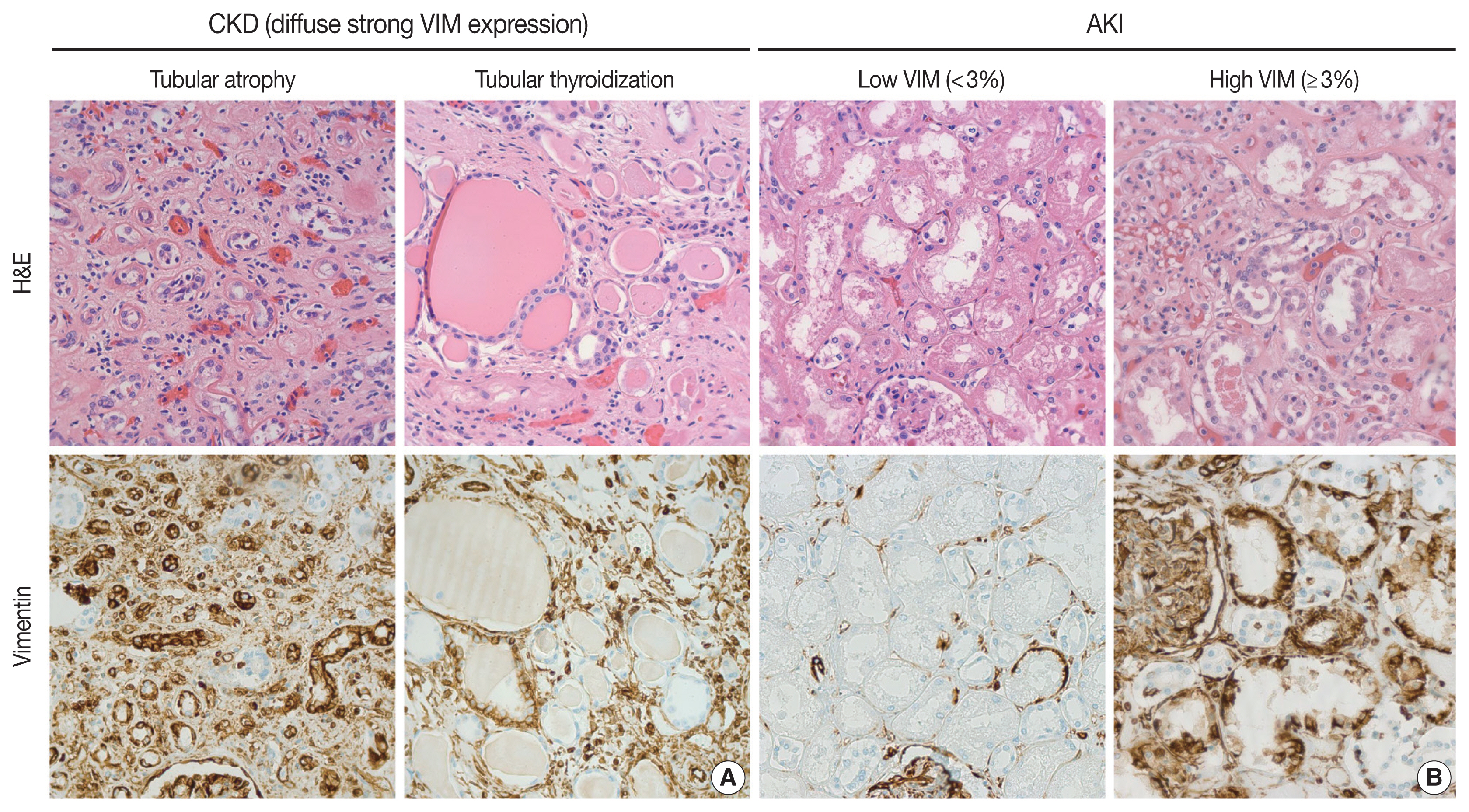
- Post-mortem assessment of vimentin expression as a biomarker for renal tubular regeneration following acute kidney injury
- Juan Carlos Alvarez Moreno, Hisham F. Bahmad, Christopher A. Febres-Aldana, Andrés Pirela, Andres Azuero, Ali Salami, Robert Poppiti
- J Pathol Transl Med. 2021;55(6):369-379. Published online October 14, 2021
- DOI: https://doi.org/10.4132/jptm.2021.08.03
- 8,767 View
- 151 Download
- 6 Web of Science
- 6 Crossref
-
Abstract
PDF
- Background
Acute kidney injury (AKI) is a common cause of morbidity and mortality. It mainly targets the renal tubular epithelium with pathological changes, referred to as acute tubular injury. The latter is followed by a regenerative response that is difficult to visualize on routine hematoxylin and eosin (H&E) stains. In this study, we examined the regenerative capacity of renal tubules by correlating vimentin (VIM) immunohistochemical (IHC) expression and pathological findings of AKI and renal tubular regeneration (RTR) on H&E.
Methods
We reviewed 23 autopsies performed in the clinical setting of AKI and RTR. VIM expression was scored in the renal cortical tubular epithelium using a statistical cutoff ≥ 3% for high expression and < 3% for low expression.
Results
Of the 23 kidney tissues examined, seven (30.4%) had low VIM expression, and 16 (69.6%) had high VIM expression. Kidney tissues with evidence of AKI and RTR had significantly higher VIM expression. Renal peritubular microenvironment features showing regenerative changes on H&E were associated with high VIM expression. In the univariate model, kidney tissues with RTR were 18-fold more likely to have high VIM expression.
Conclusions
In conclusion, our findings suggest that VIM could serve as an IHC marker for RTR following AKI. However, correlation with H&E findings remains critical to excluding chronic tubular damage. Collectively, our preliminary results pave the way for future studies including a larger sample size to validate the use of VIM as a reliable biomarker for RTR. -
Citations
Citations to this article as recorded by- Influence of Vimentin and E-cadherin on the development of interstitial fibrosis in focal and segmental glomerulosclerosis – FSGS
Laura Penna Rocha, Monise Gini Urzêdo, Nayne Isabelli Zugolaro Donzelli, Bruna de Freitas Oliveira, Crislaine Aparecida Silva, Bruna Cunha Zaidan, Régia Caroline Peixoto Lira Fusco, Marlene Antônia dos Reis, Juliana Reis Machado
Tissue and Cell.2026; 102: 103555. CrossRef - Myocardial Infarction Injury Is Exacerbated by Nicotine in Vape Aerosol Exposure
Clarissa Savko, Carolina Esquer, Claudia Molinaro, Sophie Rokaw, Abraham G. Shain, Faid Jaafar, Morgan K. Wright, Joy A. Phillips, Tyler Hopkins, Sama Mikhail, Abigail Rieder, Ariana Mardani, Barbara Bailey, Mark A. Sussman
Journal of the American Heart Association.2025;[Epub] CrossRef - Spatio-temporal transcriptomic analysis reveals distinct nephrotoxicity, DNA damage, and regeneration response after cisplatin
Lukas S. Wijaya, Steven J. Kunnen, Panuwat Trairatphisan, Ciarán P. Fisher, Meredith E. Crosby, Kai Schaefer, Karen Bodié, Erin E. Vaughan, Laura Breidenbach, Thomas Reich, Diana Clausznitzer, Sylvestre Bonnet, Sipeng Zheng, Chantal Pont, James L. Stevens
Cell Biology and Toxicology.2025;[Epub] CrossRef - Characterization of macrophages in ischemia–reperfusion injury-induced acute kidney injury based on single-cell RNA-Seq and bulk RNA-Seq analysis
Qin Wang, Yuxing Liu, Yan Zhang, Siyuan Zhang, Meifang Zhao, Zhangzhe Peng, Hui Xu, Hao Huang
International Immunopharmacology.2024; 130: 111754. CrossRef - Renal tubular necrosis associated with anagrelide administration: a case report
Atsushi Sawase, Mineaki Kitamura, Misato Morimoto, Haruka Fukuda, Tadashi Uramatsu, Eisuke Katafuchi, Hiroshi Yamashita, Toshiyuki Nakayama, Hiroshi Mukae, Tomoya Nishino
CEN Case Reports.2024; 13(6): 510. CrossRef - Morin attenuates sepsis-induced acute kidney injury by regulating inflammatory responses, oxidative stress and tubular regeneration (morin and sepsis-induced acute kidney injury)
Aya M. Shehata, Nagui H. Fares, Basma H. Amin, Asmaa A. Mahmoud, Yomna I. Mahmoud
Environmental Toxicology and Pharmacology.2024; 111: 104543. CrossRef
- Influence of Vimentin and E-cadherin on the development of interstitial fibrosis in focal and segmental glomerulosclerosis – FSGS
- Quilty Lesions in the Endomyocardial Biopsies after Heart Transplantation
- Haeyon Cho, Jin-Oh Choi, Eun-Seok Jeon, Jung-Sun Kim
- J Pathol Transl Med. 2019;53(1):50-56. Published online December 26, 2018
- DOI: https://doi.org/10.4132/jptm.2018.11.30
- 9,537 View
- 132 Download
- 7 Web of Science
- 7 Crossref
-
Abstract
PDF
 Supplementary Material
 - Background
The aim of this study was to investigate the clinical significance of Quilty lesions in endomyocardial biopsies (EMBs) of cardiac transplantation patients.
Methods
A total of 1190EMBs from 117 cardiac transplantation patients were evaluated histologically for Quilty lesions,acute cellular rejection, and antibody-mediated rejection. Cardiac allograft vasculopathy wasdiagnosed by computed tomography coronary angiography. Clinical information, including thepatients’ survival was retrieved by a review of medical records.
Results
Eighty-eight patients(75.2%) were diagnosed with Quilty lesions, which were significantly associated with acute cellularrejection, but not with acute cellular rejection ≥ 2R or antibody-mediated rejection. In patientsdiagnosed with both Quilty lesions and acute cellular rejection, the time-to-onset of Quilty lesionsfrom transplantation was longer than that of acute cellular rejections. We found a significant associationbetween Quilty lesions and cardiac allograft vasculopathy. No significant relationship wasfound between Quilty lesions and the patients’ survival.
Conclusions
Quilty lesion may be an indicator of previous acute cellular rejection rather than a predictor for future acute cellular rejection. -
Citations
Citations to this article as recorded by- A molecular reappraisal of quilty lesions: Insights from tissue and circulating biomarkers in heart transplantation
Andrea Fernandez Valledor, Cathrine M. Moeller, Adi Hertz, Daniel Oren, Ilan Richter, Boaz Elad, Julia Baranowska, Salwa Rahman, Carolyn Hennecken, Afsana Rahman, Dor Lotan, David Bae, Adil Yunis, Justin A. Fried, Ersilia M. DeFilippis, David T. Majure, J
The Journal of Heart and Lung Transplantation.2026; 45(5): 724. CrossRef - The roles of tertiary lymphoid structures in orchestrating immune responses in peripheral organs
Keisuke Taniguchi, Takahisa Yoshikawa, Motoko Yanagita
Inflammation and Regeneration.2025;[Epub] CrossRef - The human myocardium harbors a population of naive B-cells with a distinctive gene expression signature conserved across species
Kevin C. Bermea, Nicolas Kostelecky, Sylvie T. Rousseau, Chieh-Yu Lin, Luigi Adamo
Frontiers in Immunology.2022;[Epub] CrossRef - Examination of tracheal allografts after long-term survival in dogs
Tao Lu, Yiwei Huang, Yulei Qiao, Yongxing Zhang, Yu Liu
European Journal of Cardio-Thoracic Surgery.2021; 59(1): 155. CrossRef - Essentials in the diagnosis of postoperative myocardial lesions similar to or unrelated to rejection in heart transplant
Costel Dumitru, Ancuta Zazgyva, Adriana Habor, Ovidiu Cotoi, Horațiu Suciu, Carmen Cotrutz, Bogdan Grecu, Ileana Anca Sin
Revista Romana de Medicina de Laborator.2021; 29(3): 307. CrossRef - Clinical outcome of donor heart with prolonged cold ischemic time: A single‐center study
Fazal Shafiq, Yixuan Wang, Geng Li, Zongtao Liu, Fei Li, Ying Zhou, Li Xu, Xingjian Hu, Nianguo Dong
Journal of Cardiac Surgery.2020; 35(2): 397. CrossRef - The XVth Banff Conference on Allograft Pathology the Banff Workshop Heart Report: Improving the diagnostic yield from endomyocardial biopsies and Quilty effect revisited
Jean-Paul Duong Van Huyen, Marny Fedrigo, Gregory A. Fishbein, Ornella Leone, Desley Neil, Charles Marboe, Eliot Peyster, Jan von der Thüsen, Alexandre Loupy, Michael Mengel, Monica P. Revelo, Benjamin Adam, Patrick Bruneval, Annalisa Angelini, Dylan V. M
American Journal of Transplantation.2020; 20(12): 3308. CrossRef
- A molecular reappraisal of quilty lesions: Insights from tissue and circulating biomarkers in heart transplantation
Review
- Acute Atherosis of the Uterine Spiral Arteries: Clinicopathologic Implications
- Joo-Yeon Kim, Yeon Mee Kim
- J Pathol Transl Med. 2015;49(6):462-471. Published online November 4, 2015
- DOI: https://doi.org/10.4132/jptm.2015.10.23
- 22,581 View
- 246 Download
- 36 Web of Science
- 40 Crossref
-
Abstract
PDF
- Acute atherosis is unique vascular changes of the placenta associated with poor placentation. It is characterized by subendothelial lipid-filled foam cells, fibrinoid necrosis of the arterial wall, perivascular lymphocytic infiltration, and it is histologically similar to early-stage atherosclerosis. Acute atherosis is rare in normal pregnancies, but is frequently observed in non- transformed spiral arteries in abnormal pregnancies, such as preeclampsia, small for gestational age (SGA), fetal death, spontaneous preterm labor and preterm premature rupture of membranes. In preeclampsia, spiral arteries fail to develop physiologic transformation and retain thick walls and a narrow lumen. Failure of physiologic transformation of spiral arteries is believed to be the main cause of uteroplacental ischemia, which can lead to the production of anti-angiogenic factors and induce endothelial dysfunction and eventually predispose the pregnancy to preeclampsia. Acute atherosis is more frequently observed in the spiral arteries of the decidua of the placenta (parietalis or basalis) than in the decidual or myometrial segments of the placental bed. The presence and deeper location of acute atherosis is associated with poorer pregnancy outcomes, more severe disease, earlier onset of preeclampsia, and a greater frequency of SGA neonates in patients with preeclampsia. Moreover, the idea that the presence of acute atherosis in the placenta may increase the risk of future cardiovascular disease in women with a history of preeclampsia is of growing concern. Therefore, placental examination is crucial for retrospective investigation of pregnancy complications and outcomes, and accurate placental pathology based on universal diagnostic criteria in patients with abnormal pregnancies is essential for clinicopathologic correlation.
-
Citations
Citations to this article as recorded by- Placental vascular remodeling in preeclampsia: A three-dimensional analysis of microvascular alterations across disease severity
Mingqun Li, Xiaoqiang Han, Yao Peng, Yang He, Qiangqiang You, Jiaqi Zhang
Placenta.2026; 174: 96. CrossRef - Placental aberrant inflammation and spatial-specific lipid metabolism contribute to hypertensive disorder of pregnancy susceptibility in preeclampsia offspring
Pei-ran Hu, Jing-hui Xu, Yan Shi, Ying Zhu, Gao-chen Zhang, Jie-ru Yang, Yue Xu, Ming-hao Li, Xian-hua Lin, Yu Zhang, He-feng Huang
Biochimica et Biophysica Acta (BBA) - Molecular Basis of Disease.2026; 1872(4): 168168. CrossRef - Cardiac Implications of Preeclampsia: A Review
Beani J. Forst, Linda R. Chambliss, David S. Majdalany
Journal of Personalized Medicine.2026; 16(5): 265. CrossRef - Circulating vascular endothelial growth factor receptor‐3, a pro‐lymphangiogenic and pro‐angiogenic mediator, is decreased in pre‐eclampsia
Ana C. Palei, Julyane N. S. Kaihara, Ricardo C. Cavalli, Valeria C. Sandrim
International Journal of Gynecology & Obstetrics.2025; 168(1): 210. CrossRef - ECHS1 as a Lipid Metabolism Biomarker for Pediatric Focal Segmental Glomerulosclerosis
Chao He, Wei Peng, Sheng Li, Can Xu, Xiuping Chen, Yuanhan Qin, Nasar Alwahaibi
PLOS ONE.2025; 20(3): e0319049. CrossRef - PlacEntal Acute atherosis RefLecting Subclinical systemic atherosclerosis in women up to 20 years after pre-eclampsia (PEARLS): research protocol for a cohort study
Gwyneth Jansen, Robert-Jan Alers, Emma BNJ Janssen, Laura M Jorissen, Eri Morina - Shijaku, Carmen Severens-Rijvers, Arnoud van ’t Hof, J van Drongelen, Ralph R Scholten, Salwan Al-Nasiry, Droima Stevens, Wessel Ganzevoort, Sanne Gordijn, Jérôme Cornette,
BMJ Open.2025; 15(5): e100542. CrossRef - Understanding Preeclampsia: Cardiovascular Pathophysiology, Histopathological Insights and Molecular Biomarkers
Kaltrina Kutllovci Hasani, Nurxhan Ajeti, Nandu Goswami
Medical Sciences.2025; 13(3): 154. CrossRef - Evidence that atherosis of the spiral artery represents atherosclerotic lesions similar to those of native and transplant-induced atherosclerosis: implications for understanding the pathophysiology of obstetrical syndromes and long-term cardiovascular ris
Carlos A. Labarrere, Roberto Romero, Hector L. DiCarlo, James W. Hardin, Yeon Mee Kim, Arun Meyyazhagan, Offer Erez, Piya Chaemsaithong, Liliana Voto, Awoniyi Awonuga, Tinnakorn Chaiworapongsa, Ghassan S. Kassab
American Journal of Obstetrics and Gynecology.2025;[Epub] CrossRef - Human Placenta and Evolving Insights into Pathological Changes of Preeclampsia: A Comprehensive Review of the Last Decade
Diana Maria Chiorean, Esra Cobankent Aytekin, Melinda-Ildiko Mitranovici, Sabin Gligore Turdean, Mirpooya Salehi Moharer, Ovidiu Simion Cotoi, Havva Serap Toru
Fetal and Pediatric Pathology.2024; 43(1): 33. CrossRef - Effects of hypertensive disorders of pregnancy on the complications in very low birth weight neonates
Baoquan Zhang, Xiujuan Chen, Changyi Yang, Huiying Shi, Wenlong Xiu
Hypertension in Pregnancy.2024;[Epub] CrossRef - Prevention of Pregnancy Complications Using a Multimodal Lifestyle, Screening, and Medical Model
Jim Parker, Pierre Hofstee, Shaun Brennecke
Journal of Clinical Medicine.2024; 13(15): 4344. CrossRef - Placental growth factor mediates pathological uterine angiogenesis by activating the NFAT5-SGK1 signaling axis in the endometrium: implications for preeclampsia development
Janet P. Raja Xavier, Toshiyuki Okumura, Melina Apweiler, Nirzari A. Chacko, Yogesh Singh, Sara Y Brucker, Satoru Takeda, Florian Lang, Madhuri S Salker
Biological Research.2024;[Epub] CrossRef - Genome-wide DNA methylation and gene expression in human placentas derived from assisted reproductive technology
Pauliina Auvinen, Jussi Vehviläinen, Karita Rämö, Ida Laukkanen, Heidi Marjonen-Lindblad, Essi Wallén, Viveca Söderström-Anttila, Hanna Kahila, Christel Hydén-Granskog, Timo Tuuri, Aila Tiitinen, Nina Kaminen-Ahola
Communications Medicine.2024;[Epub] CrossRef - Missing links in preeclampsia cell model systems of endothelial dysfunction
Sarah Viana-Mattioli, Miriam Helena Fonseca-Alaniz, Iguaracy Pinheiro-de-Sousa, José Eduardo Krieger, Valéria Cristina Sandrim
Trends in Molecular Medicine.2023; 29(7): 541. CrossRef - Roles of maternal HDL during pregnancy
Laura A. Woollett, Janet M. Catov, Helen N. Jones
Biochimica et Biophysica Acta (BBA) - Molecular and Cell Biology of Lipids.2022; 1867(3): 159106. CrossRef - The role of the placenta in spontaneous preterm labor and delivery with intact membranes
Sunil Jaiman, Roberto Romero, Gaurav Bhatti, Eunjung Jung, Francesca Gotsch, Manaphat Suksai, Dahiana M. Gallo, Tinnakorn Chaiworapongsa, Nicholas Kadar
Journal of Perinatal Medicine.2022; 50(5): 553. CrossRef - Gestational Antibodies to C. pneumoniae, H. pylori and CMV in Women with Preeclampsia and in Matched Controls
Abdul Wajid, David Todem, Mark R. Schleiss, David F. Colombo, Nigel S. Paneth
Maternal and Child Health Journal.2022; 26(10): 2040. CrossRef - Toward a new taxonomy of obstetrical disease: improved performance of maternal blood biomarkers for the great obstetrical syndromes when classified according to placental pathology
Roberto Romero, Eunjung Jung, Tinnakorn Chaiworapongsa, Offer Erez, Dereje W. Gudicha, Yeon Mee Kim, Jung-Sun Kim, Bomi Kim, Juan Pedro Kusanovic, Francesca Gotsch, Andreea B. Taran, Bo Hyun Yoon, Sonia S. Hassan, Chaur-Dong Hsu, Piya Chaemsaithong, Nardh
American Journal of Obstetrics and Gynecology.2022; 227(4): 615.e1. CrossRef - Preeclampsia and Fetal Growth Restriction as Risk Factors of Future Maternal Cardiovascular Disease—A Review
Sylwia Sławek-Szmyt, Katarzyna Kawka-Paciorkowska, Aleksandra Ciepłucha, Maciej Lesiak, Mariola Ropacka-Lesiak
Journal of Clinical Medicine.2022; 11(20): 6048. CrossRef - The Role of NF-κB in Uterine Spiral Arteries Remodeling, Insight into the Cornerstone of Preeclampsia
Maciej W. Socha, Bartosz Malinowski, Oskar Puk, Mateusz Wartęga, Martyna Stankiewicz, Anita Kazdepka-Ziemińska, Michał Wiciński
International Journal of Molecular Sciences.2021; 22(2): 704. CrossRef - Pathogenesis of uteroplacental acute atherosis: An update on current research
Shu Li, Yan‐Wei Hu
American Journal of Reproductive Immunology.2021;[Epub] CrossRef - Disorders of placental villous maturation are present in one-third of cases with spontaneous preterm labor
Sunil Jaiman, Roberto Romero, Percy Pacora, Offer Erez, Eunjung Jung, Adi L. Tarca, Gaurav Bhatti, Lami Yeo, Yeon Mee Kim, Chong Jai Kim, Jung-Sun Kim, Faisal Qureshi, Suzanne M. Jacques, Nardhy Gomez-Lopez, Chaur-Dong Hsu
Journal of Perinatal Medicine.2021; 49(4): 412. CrossRef - The COVID-19 Pandemic: an Appraisal of its Impact on Human Immunodeficiency Virus Infection and Pre-Eclampsia
Rowen Govender, Jagidesa Moodley, Thajasvarie Naicker
Current Hypertension Reports.2021;[Epub] CrossRef - Acute Atherosis Lesions at the Fetal-Maternal Border: Current Knowledge and Implications for Maternal Cardiovascular Health
Daniel Pitz Jacobsen, Heidi Elisabeth Fjeldstad, Guro Mørk Johnsen, Ingrid Knutsdotter Fosheim, Kjartan Moe, Patji Alnæs-Katjavivi, Ralf Dechend, Meryam Sugulle, Anne Cathrine Staff
Frontiers in Immunology.2021;[Epub] CrossRef - Aetiology, prophylaxis and management of preeclampsia
Karolina Gronkowska
Acta Universitatis Lodziensis. Folia Biologica et Oecologica.2021; 17: 111. CrossRef - HMOX1 is partly responsible for phenotypic and functional abnormalities in mesenchymal stem cells/stromal cells from placenta of preeclampsia (PE) patients
Yasser S. Basmaeil, Dana Algudiri, Reem Alenzi, Abdullah Al Subayyil, Ayodele Alaiya, Tanvir Khatlani
Stem Cell Research & Therapy.2020;[Epub] CrossRef - Analyzing Preeclampsia as the Tip of the Iceberg Represented by Women with Long-Term Cardiovascular Disease, Atherosclerosis, and Inflammation
Angélica Lemos Debs Diniz, Maria Marta Bini Martins Paes, Aline Debs Diniz
Current Atherosclerosis Reports.2020;[Epub] CrossRef - Lipids in preeclampsia: pathogenic parallels to atherosclerosis
V. I. Shcherbakov, Ya. V. Polonskaya, E. V. Kashtanova, A. V. Shirinskaya
"Arterial’naya Gipertenziya" ("Arterial Hypertension").2020; 26(2): 163. CrossRef - Transthyretin increases migration and invasion of rat placental trophoblast cells
Xiao‐Peng Ma, Chong‐Dong Liu, Guang‐Ming Cao, Zhen‐Yu Zhang
FEBS Open Bio.2020; 10(8): 1568. CrossRef - Early Onset Preeclampsia Is Associated With Glycocalyx Degradation and Reduced Microvascular Perfusion
Tracey L. Weissgerber, Oscar Garcia‐Valencia, Natasa M. Milic, Elizabeth Codsi, Hajrunisa Cubro, Meryl C. Nath, Wendy M. White, Karl A. Nath, Vesna D. Garovic
Journal of the American Heart Association.2019;[Epub] CrossRef - The immunophenotype of decidual macrophages in acute atherosis
Navleen Gill, Yaozhu Leng, Roberto Romero, Yi Xu, Bogdan Panaitescu, Derek Miller, Afrah Arif, Salma Mumuni, Faisal Qureshi, Chaur‐Dong Hsu, Sonia S. Hassan, Anne Cathrine Staff, Nardhy Gomez‐Lopez
American Journal of Reproductive Immunology.2019;[Epub] CrossRef - The potential effects of pomegranate peel extract and bee venom in improving the diabetes induced damaging of spiral artery
HIH El-Sayyad, HA El-Ghawet, AMA El-Sayed
Studies on Stem Cells Research and Therapy.2019; 5(1): 007. CrossRef - Race and risk of maternal vascular malperfusion lesions in the placenta
Vanessa Assibey-Mensah, W. Tony Parks, Alison D. Gernand, Janet M. Catov
Placenta.2018; 69: 102. CrossRef - Preclinical atherosclerosis at the time of pre‐eclamptic pregnancy and up to 10 years postpartum: systematic review and meta‐analysis
N. M. Milic, J. Milin‐Lazovic, T. L. Weissgerber, G. Trajkovic, W. M. White, V. D. Garovic
Ultrasound in Obstetrics & Gynecology.2017; 49(1): 110. CrossRef - Is an episode of suspected preterm labor that subsequently leads to a term delivery benign?
Roberto Romero, Offer Erez, Eli Maymon, Percy Pacora
American Journal of Obstetrics and Gynecology.2017; 216(2): 89. CrossRef - Establishment of the Human Uteroplacental Circulation: A Historical Perspective
Kenna Degner, Ronald R. Magness, Dinesh M. Shah
Reproductive Sciences.2017; 24(5): 753. CrossRef - Preeclampsia and coronary plaque erosion: Manifestations of endothelial dysfunction resulting in cardiovascular events in women
Saskia C.A. de Jager, John A.L. Meeuwsen, Freeke M. van Pijpen, Gerbrand A. Zoet, Arjan D. Barendrecht, Arie Franx, Gerard Pasterkamp, Bas B. van Rijn, Marie-José Goumans, Hester M. den Ruijter
European Journal of Pharmacology.2017; 816: 129. CrossRef - Placental histopathology lesions and pregnancy outcome in pregnancies complicated with symptomatic vs. non-symptomatic placenta previa
Eran Weiner, Hadas Miremberg, Ehud Grinstein, Letizia Schreiber, Shimon Ginath, Jacob Bar, Michal Kovo
Early Human Development.2016; 101: 85. CrossRef - Porphyromonas gingivalis within Placental Villous Mesenchyme and Umbilical Cord Stroma Is Associated with Adverse Pregnancy Outcome
Sizzle F. Vanterpool, Jasper V. Been, Michiel L. Houben, Peter G. J. Nikkels, Ronald R. De Krijger, Luc J. I. Zimmermann, Boris W. Kramer, Ann Progulske-Fox, Leticia Reyes, Motohiro Komaki
PLOS ONE.2016; 11(1): e0146157. CrossRef - Pregnant women with heart disease: Placental characteristics and their association with fetal adverse events
Fabio V. Lima, Paraskevi Koutrolou-Sotiropoulou, Puja B. Parikh, Cecilia Avila, Javed Butler, Kathleen Stergiopoulos
Acute Cardiac Care.2016; 18(3): 56. CrossRef
- Placental vascular remodeling in preeclampsia: A three-dimensional analysis of microvascular alterations across disease severity
Case Report
- Acute Renal Failure Associated with Gross Hematuria in a Patient with Focal Glomerulonephritis.
- Hee Jung Kim, Hyeon Joo Jeong, Dae Suk Han
- Korean J Pathol. 1997;31(3):263-268.
- 2,617 View
- 18 Download
-
Abstract
PDF
- A 58-year-old female with an episode of gross hematuria two months before and fever and chill for the past three days presented oliguric acute renal failure. She has taken NSAID intermittently for 18 years due to rheumatoid arthritis, and herb medicine for one week two months ago when gross hematuria developed. Physical examination revealed mild tenderness on costovertebral angles. Her blood pressure was 170/100 mmHg, the urinalysis showed >300 mg protein with many RBCs and 10-20 WBCs and the serum creatinine was 5.8 mg/dl. A renal biopsy performed on the 4th hospital day showed that it was overwhelmed by severe tubular lesions which reveal intratubular obstruction by massive erythrocyte casts and tubular necrosis. The glomeruli showed focal minimal crescents with many red blood cells entrapped in the crescents and in the capillaries. Immune deposits were not present. A renal failure resolved spontaneously and the patient was discharged three weeks later with creatinine of 2.4 mg/dl. In this patient, acute renal failure was considered to be due to a tubular lesion related to the glomerular bleeding from focal glomerulonephritis revealing minimal crescents.
Original Article
- Pathological Analysis of Post-Transplantation Endomyocardial Biopsies.
- Jaegul Chung, Soonae Oak, Gheeyoung Choe, Gyungyub Gong, Jooryung Huh, Eunsil Yu, Inchul Lee, Meong Gun Song, Kwang Hyun Sohn, Jae Joong Kim, Jong Goo Lee
- Korean J Pathol. 1995;29(4):431-441.
- 2,470 View
- 17 Download
-
Abstract
PDF
- Heart transplantation was first performed in 1967. It is now regarded as a well-established treatment modality for end-stage cardiac diseases. Once the transplantation is performed, endomyocardial biopsy(EMB) is the examination of choice in monitoring the transplanted heart. We analyzed the pathological findings of follow-up EMB of 6 heart transplant patients. All patients have been suffered from severe heart failure. Four patients were adult male and two were adult females. All the hearts, except for one, displayed characteristic features of dilated cardiomyopathy. The remaining heart was diagnosed as having giant cell myocarditis. Post-transplantion EMBs were performed according to the protocol and standard cardiac biopsy grading of ISHT (1990). The standards were applied for grading of cellular rejection. In five patients, there were one or two episodes of biopsy proven acute rejection, grade II or IIIA without any clinical symptoms of rejection. Immediate "pulse therapy" was performed and follow-up biopsies were done. All episodes of rejection were cleared in subsequent biopsies. All patients are doing well without evidence of cardiac problem. The postoperative monitoring of acute rejection is critical since clinical signs of rejection are usually absent. At present, EMB is regarded as the most reliable method for diagnosis and grading of acute rejection and is an efficient guide to the monitoring of the cardiac recipients. Our experience of post-transplantation EMB corresponds with previously published reports.
Case Reports
- Acute Interstitial Pneumonia (Hamman-Rich Syndrome): An Autopsy Case.
- Han Kyeom Kim, Ae Ree Kim, Min Ji Jeoung, Won Hee Seo, Jee yeoun Lee, Su Hyun Park
- Korean J Pathol. 1997;31(4):366-374.
- 2,904 View
- 41 Download
-
Abstract
PDF
- Acute interstitial pneumonia is a fulminant disease of unknown etiology that usually occurs in a previously healthy person and produces the histologic findings of the organizing phase of diffuse alveolar damage. We experienced an autopsy case of acute interstitial pneumonia of unknown etiology. The patient was a 48 year old man who had been healthy and had not been exposed to organic dusts or other toxic materials. The chief complaints represented were dyspnea and a dry cough for several weeks before hospitalization, and the chest radiographs showed bilateral interstitial infiltrates. Patchy consolidation of air space was also identified and ground-glass attenuation similar to those described in ARDS was detected on high-resolution computed tomography. Steroid pulse therapy, mechanical ventilation, and antibiotics for superimposed bacterial infection were performed, but the symptoms did not improve and the patient died of generalized respiratory insufficiency and severe hypoxemia 2 1/2 months after hospitalization. At autopsy the macroscopic and microscopic findings were confined mainly to the lungs. On the whole, both lungs were firm in consistency and the external surface showed a cobblestone appearance. The cut surface showed almost complete replacement of the normal lung parenchyma with gray to yellow fibrous tissue with a little residual functional area remaining. The pathology of both open lung biopsy and autopsy tissue showed marked hyperplasia of type II pneumocytes, hyaline membrane formation, thickening of the alveolar wall due to extensive fibroblast proliferation, and relatively abundant young collagen deposition in the interstitium. An immunohistochemical stain for cytokeratin revealed epithelial hyperplasia and showed that the alveolar spaces were markedly shrunken by fibrous tissue.
- An Unusual Type of Acute Renal Failure due to Extensive Crystal Deposition in the Renal Tubular Epithelium and Interstitium: A Case Report.
- Ja Seung Koo, Eunah Shin, Shin Woo Kang, Hyeon Joo Jeong
- Korean J Pathol. 2004;38(5):337-340.
- 2,249 View
- 19 Download
-
Abstract
PDF
- Acute tubular necrosis is a major cause of acute renal failure. Acute renal failure that is caused by crystal deposition can result from drug toxicity, lymphoplasmacytic neoplasms, ingestion of industrial organic solvents, or intratubular obstruction due to degenerated red blood cells and red blood cell casts. We herein present an uncommon case of acute renal failure in a 57-year-old woman showing an unusually massive accumulation of variable-sized, round, ellipsoid or rhomboid, pale-pink, refractile bodies in the proximal and distal tubular epithelial cells, interstitial macrophages and Bowman's spaces. These bodies were electron dense with a maximum diameter of 3 micrometer. The information we gathered from the patient history, the laboratory data and the various histochemical and immunohistochemical analyses failed to reveal the exact nature of these crystal-like structures.
Original Article
- Application of Immunohistochemical Stain for Granulocytic Sarcoma.
- Yeong Ju Woo, Chan Hwan Kim, Jong Eun Joo
- Korean J Pathol. 1994;28(1):30-37.
- 2,461 View
- 28 Download
-
Abstract
PDF
- Granulocytic sarcoma is a rare localized tumor composed of granulocytic precusor cells. Granu-locytic sarcoma occurs in a variety of clinical conditions and it is often misdiagnosed histologically. Differential diagnosis frorh lymphoma or nonhematopoietic malignancies such as undifferentiated carcinoma or sarcoma is difficult in the routing histologic examination. An evaluation of clinical and histopathologic features was done on 4 cases of granulocytic sarcoma which were diagnosed at Pusan Paik Hospital from 1988 to 1992. During the period, 282 cases of myelogenous leukemia were diagnosed. Immunohistochemical reaction for lysozyme, myelopero-xidase, leukocyte common antigen, epthelial membrane antigen and cytokeratin was assessed comparing to lymphoma and undifferentiated carcinoma. The histologic features of the granulocytic sarcoma revealed thin nuclear membrane, fine chromatin pattern and one or two small nucleoli. It also often involved the vascular wall and infiltrated the native structures without destruction. Immunohistochemical stain revealed that all(4 cases) of granulocytic sarcoma showed diffuse and strong positivity for myeloperoxidase, and partial but strong positivity for lysozyme. One case of granulocytic sarcoma was negative and 3 cases revealed focal positive reaction for LCA, and all 4 cases was negative for cytokeratin and EMA. In summary, careful observation under light microscopy with immunohistochemical stain for myeloperoxidase, lysozyme, and LCA is helpful in the differential diagnosis of granulocytic sarcoma from malignant lymphoma and cytokeratin and EMA is useful for differential diagnosis from undifferentiated carcinoma.
Case Report
- Hepatic Candidiasis: A case occurred in a patient with leukemia.
- Chan Il Park, Sun Hee Sung, Eun Kyung Han, Ho Guen Kim
- Korean J Pathol. 1991;25(3):275-277.
- 2,239 View
- 25 Download
-
Abstract
PDF
- In view of the possible role of portal circulation in hematogenous spread of Candida species, a case of hepatic candidiasis occurred in an eight-year-old child with acute lymphoblastic leukemia (ALL) treated by chemotherapy is presented. Symptoms and signs referable to the hepatic disease in this patient included hepatomegaly, icteric sclera and abdominal pain. There were no particular manifestations suggestive of deep mycotic involvement of any sepcific organs or tissues other than the liver. Culture of the blood was negative for one month. On the 24th hospital day the patient died with the presumptive diagnosis of ALL, disseminated intravascular coagulation, acute renal failure, pulmonary edema, cholecystitis and oral thrush. A needle necropsy was performed and revealed fungal aggregates replacing the large foci of hepatic cell loss. It is suggested that, when the gastrointestinal tract serves as the portal of entry, the liver could be the visceral organ involved first in the course of disseminated candidiasis.
Original Articles
- Pathologic Analysis of 2159 Cases of Appendix.
- Chan Sik Park, Mee Soo Chang, In Ae Park, Yong Il Kim, Gheeyoung Choe
- Korean J Pathol. 2000;34(1):39-49.
- 3,009 View
- 49 Download
-
Abstract
PDF
- We reviewed 2159 consecutive cases of surgically resected appendices. The appendectomy specimen consisted of 91 cases of acute focal appendicitis (5.4%), 926 cases of acute suppurative appendicitis (55.1%), 228 cases of acute gangrenous appendicitis (13.6%), 63 cases of periappendicitis (3.8%), 13 cases of pure fibrous obliteration of the lumen (FOL; 0.8%), 18 cases of other diseases (7 mucoceles, 2 mucinous cystic neoplasms, 4 carcinoids, 2 metastatic carcinomas, 2 tuberculous appendicitides, and 1 eosinophilic appendicitis; 1%), and 342 cases with no diagnostic abnormality (20.3%). Patients having acute appendicitis ranged from 3 to 84 years of age, and patients in their 10's and 20's occupied over half of 2159 cases. Diagnostic accuracy of the acute appendicitis was 79.7%. Incidence of the acute appendicitis was suspected to be 7.2/100,000/year. Twenty eight cases of acute appendicitis were associated with diverticula. In the former acute primary diverticulitis led to acute appendicitis in 14 of 28 cases. Among 478 incidental appendectomy cases, there were 3 acute focal appendicitides, 1 acute suppurative appendicitis, 1 eosinophilic appendicitis, 32 periappendicitides, 1 mucocele, 40 pure FOLs, 1 deciduosis, 1 endometriosis, and 1 diverticulosis without inflammation. There were 69 cases of FOL (32 complete forms and 37 incomplete forms), among which 13 cases were associated with acute appendicitis. FOL was more frequent in female patients as well as patients over 40 years of age. Incomplete FOL was considered to progress to complete form with age. The incidence of appendiceal diverticula was higher, whereas the incidences of carcinoid tumor and FOL were lower compared with that in the western report. In 14 of 28 cases the appendiceal diverticulum was the site in which acute appendicitis began.
- Apoptosis and Cell Proliferation in Experimental Acute Tubular Necrosis Induced by Intramuscular Glycerol Injection.
- Wan Seop Kim, Jung Woo Noh, Moon Hyang Park
- Korean J Pathol. 2003;37(1):41-49.
- 2,607 View
- 18 Download
-
Abstract
PDF
- BACKGROUND
Acute tubular necrosis (ATN) is the most common cause of acute renal failure. It is characterized by the destruction of tubular epithelial cells. To examine apoptosis and proliferative activity of tubular cells in the course of acute tubular necrosis, we induced acute renal failure by intramuscular hypertonic glycerol injection to New Zealand White rabbits.
METHODS
The immunohistochemistry was done for Ki-67 and tissue-transglutaminase (tTG), and the terminal deoxynucleotidyl transferase mediated nick end labeling (TUNEL) method was performed using a total of 77 renal specimens including 29 gun biopsies and 48 nephrectomiy specimens.
RESULTS
Widespread tubular injury with pigment casts and interstitial hemorrhage were noted. The tubular proliferation index was increased at 2 hours after glycerol injection, and the index peaked at 3 hours. The second cell proliferation peak was noted at 3 days. Apoptotic cells were identified by TUNEL and tTG staining. The apoptotic index was significantly increased, and it peaked at 24 hours after glycerol injection. There was a significant correlation between the proliferation index (MIB-1) the and the apototic index (TUNEL)(p= 0.001). A DNA ladder pattern was observed at 6 to 8 hours.
CONCLUSIONS
Tubular cell proliferation and apoptosis occur in the early phase after the induction of acute tubular necrosis, and the excess hyperplastic epithelial cells appear to be eliminated by apoptosis.
- Clinicopathologic Characteristics of Ulcerative Colitis Diagnosed by Endoscopic Biopsy Specimen: An analysis of discrepancy between clinical and pathologic diagnosis.
- Jong Yup Bae, Ho Guen Kim
- Korean J Pathol. 1996;30(12):1091-1098.
- 2,302 View
- 16 Download
-
Abstract
PDF
- Chronic ulcerative colitis is a systemic inflammatory disease with uncertain etiology primarily involving the colonic mucosa. The mucosal biopsy interpretation is important for an evaluation of the disease state and further medical or surgical treatment. However, few clinical and pathological studies of the endoscopic diagnosis of this disease are available in Korea. Therefore, we evaluated the clinical and pathological characteristics of it diagnosed by endoscopic biopsy and analysed the reasons for the discrepancy between clinical and pathologic diagnosis for a more accurate endoscopic mucosal biopsy diagnosis in the future. A total of 702 cases of colonic mucosal biopsy specimens during Feb. 1994 and Jan. 1995 at Severance hospital, Yonsei University College of Medicine were reevaluated for the study. A clinical diagnosis of ulcerative colitis, after endoscopic examination, was made in 61(8.7%) cases. A pathological diagnosis was made when there is an increased inflammatory cell infiltration in the mucosa with evidences of a chronic crypt injury in the biopsy specimens. Using this criteria, a diagnosis was made in 32(52.3%) cases. In 29 cases the diagnosis was made in the first biopsy specimen and in the remaining 3 cases the diagnosis was made in the second or third biopsy specimens. No pathologic diagnosis of ulcerative colitis was made in the cases that clinical diagnosis was not. In the 32 cases diagnosed as ulcerative colitis, 14 cases were involved the rectum and sigmoid colon, 9 cases were involved up to the descending colon, 1 case was involved up to the transverse colon and 8 cases showed pancolonic involvement. In 29 cases, which ulcerative colitis was suspected clinically but was not consistent with it pathologically, 8 cases were proved to be ischemic colitis, 5 cases were acute infectious colitis and one case was Crohn's disease by repeat examination and follow up. Ten cases were histologically within normal range and lesions subsided spontaneously with no recurrence. A conclusive diagnosis could not be made in 5 cases during this study period. From these results, we conclude that ulcerative colitis can be diagnosed accurately by endoscopic biopsy, and clinical follow up and repeat examination are valuable in the differential diagnosis of this disease.
- Incidence of Acute Placental Inflammation through Histopathological Analysis: One year experience in 1995 at Seoul National University Hospital.
- Hyun Ju Yoo, Yun Kyung Kang, Chong Jai Kim, Jung Sun Kim, Tae Sook Kim, Kyung Cheun Jung, Kyo Hoon Park, Jong Kwan Jun, Bo Hyun Yoon
- Korean J Pathol. 1996;30(12):1123-1128.
- 2,272 View
- 20 Download
-
Abstract
PDF
- The diagnosis of acute inflammation of the placenta, represented as acute chorioamnionitis, is important in that it is associated with a poor clinical outcome for both the mother and the fetus, including major perinatal morbidities such as sepsis, respiratory distress syndrome, and CNS damage. However, current medical trends in Korea seem to overlook the significance of a histopathological diagnosis of acute placental inflammation, mainly due to the indifferences of clinicians and pathologists. Since late 1993, histopathological examinations have been performed on preterm placentas at Seoul National University. These examinations have demonstrated acute placental inflammation in a significant number of cases. In the present study the incidence of acute placental inflammation was analyzed in 521 placentas which were submitted for pathological examinations in 1995. Examinations were performed to provide basic information on the incidence and profile of acute placental inflammation in this hospital and, thereby, to emphasize the significance of histopathological examinations of the placenta in the routine surgical pathology service. Among the 521 placentas, acute inflammation was found in 194 cases (37.2%). In preterm placentas acute inflammation was found in 39.6% of the cases (67/169), while 36.1% (127/352) of term placentas showed acute inflammation. Taking the delivery mode into account, 26.3% (49/186) of the placentas delivered by cesarean section showed acute inflammation, while 43.3% (145/335) of the transvaginally delivered placentas showed inflammation. The present analysis demonstrates the existence of acute inflammation in a significant proportion of placentas with different clinical settings. The importance of a histopathological examination in routine hospital practice should be emphasized.
- Correlation between Renal Growth Retardation and Apoptosis of Cortical Tubules in Experimentally Induced Acute Ascending Pyelonephritis in Infant Rat.
- Sun Hee Sung, Soyoun Woo, Seung Joo Lee
- Korean J Pathol. 2000;34(12):1001-1008.
- 2,098 View
- 15 Download
-
Abstract
PDF
- The infant kidney is more vulnerable to infections than the adult kidney. It is common that acute pyelonephritis (APN) during infancy and early childhood manifests growth retardation of kidney, ultimately leading to chronic renal failure. However, little is known about the pathogenesis of renal growth retardation in APN in youth. To understand the mechanism underlying the cortical lesions, urinary tract infection was induced in infant rats. To induce ascending APN, saline solution containing Escherichia coli (ATCC No. 25922) 107 bacteria/ml was infused into the bladder through the 16 gage silicone cannula in three-week-old weaning Sprague Dawley rats (weight 50~60 g, n=66). In the normal control group (n=20), saline was infused. Experimental groups were divided according to the treatment into the APN group (APN without any treatment, n=23) and TRX group (APN with ceftriaxone treatment, n=23). After performing the histopathologic examination, including inflammatory score, fibrosis score, and tubular atrophy score, we measured the apoptosis index in the tubular cells of noninflammatory cortical area at post-infection week 1 and 3 by the in situ TUNEL method. Kidney weight was significantly decreased in the APN group compared with the normal group at postinfection week 1 and 3. In the APN group, tubulointerstitial inflammation with heavy neutrophilic infiltration was found mainly in the upper and lower poles of the kidney in both the first and third week groups. Fibrosis was dominant in the third week of the APN group. However, inflammation and fibrosis were not significantly improved by TRX treatment. The apoptotic index of tubular cells was significantly increased in noninflammatory cortical area in the first week of both APN and TRX groups. It decreased near the normal control value in the third week. TGF-beta1 protein expression was localized in the inflammatory area. There was no TGF-beta1 expression in the tubules of the noninflammatory area. These findings suggest that renal growth retardation in experimentally induced APN in infant rats is related not only with the inflammatory reaction itself but also with the increased apoptosis of tubular cells in noninflammatory area. Ceftriaxone alone does not eliminate the inflammation nor prevent growth retardation effectively.
 First
First Prev
Prev


