 E-submission
Previous issues
- Page Path
- HOME > Articles and issues > Previous issues
- Volume 55(3); May 2021
-


Reviews
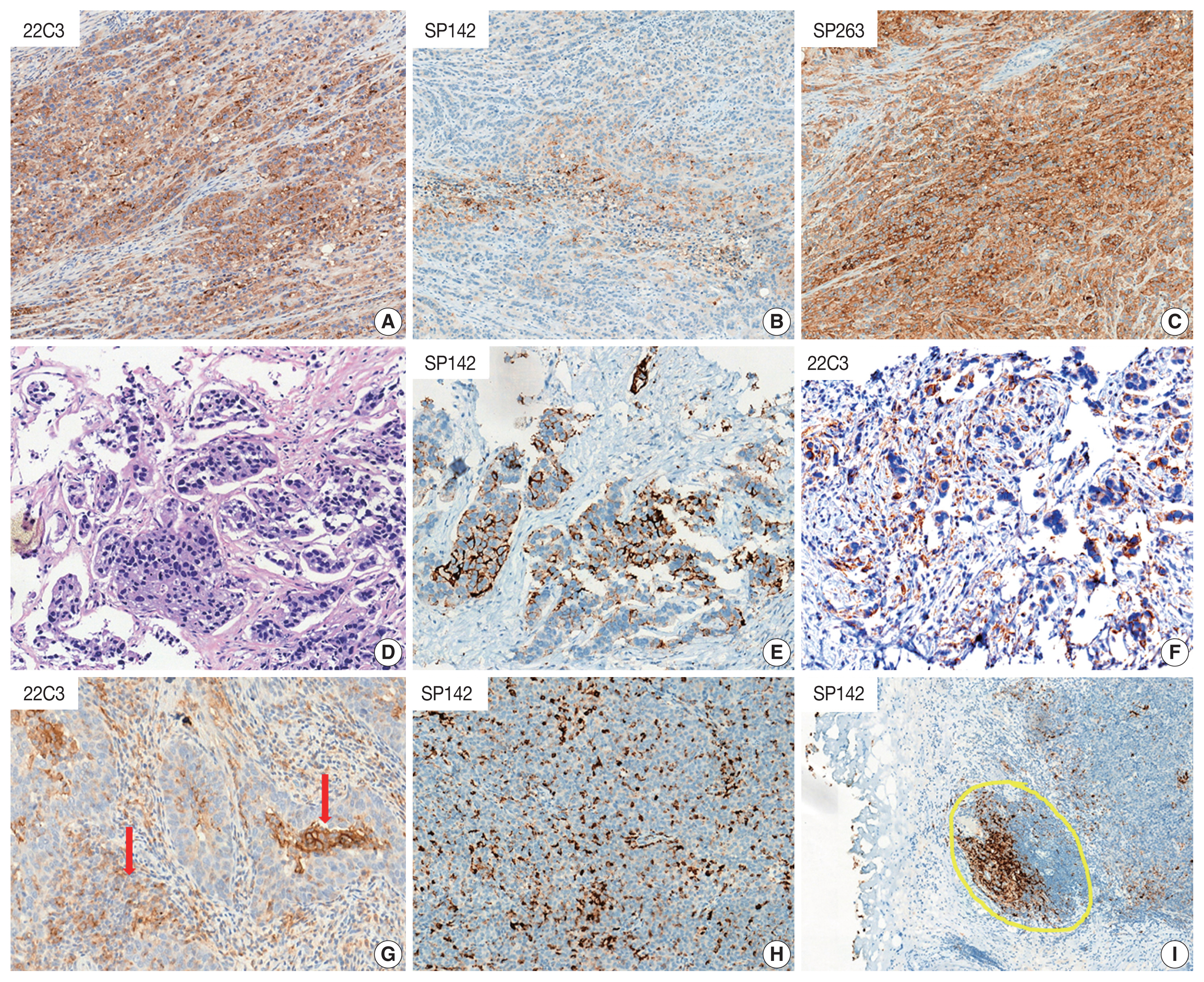
- Programmed cell death-ligand 1 assessment in urothelial carcinoma: prospect and limitation
- Kyu Sang Lee, Gheeyoung Choe
- J Pathol Transl Med. 2021;55(3):163-170. Published online April 7, 2021
- DOI: https://doi.org/10.4132/jptm.2021.02.22
- 4,882 View
- 163 Download
- 6 Web of Science
- 6 Crossref
-
 Abstract
 PDF
PDF - Programmed cell death protein 1/programmed death-ligand 1 (PD-1/PD-L1) inhibition has revolutionized the treatment paradigm of urothelial carcinoma (UC). Several PD-L1 assays are conducted to formulate appropriate treatment decisions for PD-1/PD-L1 target therapy in UC. However, each assay has its own specific requirement of antibody clones, staining platforms, scoring algorithms, and cutoffs for the determination of PD-L1 status. These prove to be challenging constraints to pathology laboratories and pathologists. Thus, the present article comprehensively demonstrates the scoring algorithm used and differences observed in each assay (22C3, SP142, and SP263). Interestingly, the SP142 score algorithm considers only immune cells and not tumor cells (TCs). It remains controversial whether SP142 expressed only in TCs truly accounts for a negative PD-L1 case. Moreover, the scoring algorithm of each assay is complex and divergent, which can result in inter-observer heterogeneity. In this regard, the development of artificial intelligence for providing assistance to pathologists in obtaining more accurate and objective results has been actively researched. To facilitate efficiency of PD-L1 testing, several previous studies attempted to integrate and harmonize each assay in UC. The performance comparison of the various PD-L1 assays demonstrated in previous studies was encouraging, the exceptional concordance rate reported between 22C3 and SP263. Although these two assays may be used interchangeably, a clinically validated algorithm for each agent must be applied.
-
Citations
Citations to this article as recorded by
- Comparison of tissue biomarkers between non-schistosoma and schistosoma-associated urothelial carcinoma
Nashwah Samir AlHariry, Enas A. El Saftawy, Basma Emad Aboulhoda, Ahmed H. Abozamel, Mansour A. Alghamdi, Amany E. Hamoud, Walaa Abd Elgawad Khalil Ghanam
Tissue and Cell.2024; 88: 102416. CrossRef - Aspectos prácticos sobre la determinación de PD-L1 en el tratamiento de carcinoma urotelial. Consenso del grupo de uropatología de la SEAP
Antonio López-Beltrán, Pilar González-Peramato, Julián Sanz-Ortega, Juan Daniel Prieto Cuadra, Isabel Trias, Rafael J. Luque Barona, María Eugenia Semidey, Pablo Maroto, Ferran Algaba
Revista Española de Patología.2023; 56(4): 261. CrossRef - Systemic treatment of advanced and metastatic urothelial cancer: The landscape in Australia
Howard Gurney, Timothy D. Clay, Niara Oliveira, Shirley Wong, Ben Tran, Carole Harris
Asia-Pacific Journal of Clinical Oncology.2023; 19(6): 585. CrossRef - PD-L1 Testing in Urothelial Carcinoma: Analysis of a Series of 1401 Cases Using Both the 22C3 and SP142 Assays
Harriet Evans, Brendan O’Sullivan, Frances Hughes, Kathryn Charles, Lee Robertson, Philippe Taniere, Salvador Diaz-Cano
Pathology and Oncology Research.2022;[Epub] CrossRef - Insights on recent innovations in bladder cancer immunotherapy
Mohamed A. Abd El‐Salam, Claire E.P. Smith, Chong‐Xian Pan
Cancer Cytopathology.2022; 130(9): 667. CrossRef - What Do We Have to Know about PD-L1 Expression in Prostate Cancer? A Systematic Literature Review. Part 1: Focus on Immunohistochemical Results with Discussion of Pre-Analytical and Interpretation Variables
Andrea Palicelli, Martina Bonacini, Stefania Croci, Cristina Magi-Galluzzi, Sofia Cañete-Portillo, Alcides Chaux, Alessandra Bisagni, Eleonora Zanetti, Dario De Biase, Beatrice Melli, Francesca Sanguedolce, Moira Ragazzi, Maria Paola Bonasoni, Alessandra
Cells.2021; 10(11): 3166. CrossRef
- Comparison of tissue biomarkers between non-schistosoma and schistosoma-associated urothelial carcinoma
- Hepatocellular adenomas: recent updates
- Haeryoung Kim, Young Nyun Park
- J Pathol Transl Med. 2021;55(3):171-180. Published online April 7, 2021
- DOI: https://doi.org/10.4132/jptm.2021.02.27
- 8,029 View
- 483 Download
- 8 Web of Science
- 9 Crossref
-
Abstract
PDF
- Hepatocellular adenoma (HCA) is a heterogeneous entity, from both the histomorphological and molecular aspects, and the resultant subclassification has brought a strong translational impact for both pathologists and clinicians. In this review, we provide an overview of the recent updates on HCA from the pathologists’ perspective and discuss several practical issues and pitfalls that may be useful for diagnostic practice.
-
Citations
Citations to this article as recorded by- Prognostic role of selection criteria for liver transplantation in patients with hepatocellular carcinoma: Review and bibliometric
Pamela Scarlett Espinoza Loyola, Diana Laura Muratalla Bautista, Karen Adela Hernández Bautista, Elizabeth Gil White, José Antonio González Moreno, Daniel Angel Torres del Real, Víctor Manuel Páez Zayas, Carla Escorza-Molina, Fernando Mondragón Rodríguez,
iLIVER.2024; 3(1): 100077. CrossRef - ACG Clinical Guideline: Focal Liver Lesions
Catherine Frenette, Mishal Mendiratta-Lala, Reena Salgia, Robert J. Wong, Bryan G. Sauer, Anjana Pillai
American Journal of Gastroenterology.2024; 119(7): 1235. CrossRef - Hepatocellular adenoma update: diagnosis, molecular classification, and clinical course
Sarah Poetter-Lang, Ahmed Ba-Ssalamah, Nina Bastati, Sami A Ba-Ssalamah, Jacqueline C Hodge, Giuseppe Brancatelli, Valérie Paradis, Valérie Vilgrain
British Journal of Radiology.2024; 97(1163): 1740. CrossRef - Fatal rupture of hepatic adenomatosis: Autopsy case and review of the literature
Sarra Ben Abderrahim, Khouloud Chérif, Zeineb Nfikha, Sarra Gharsallaoui, Imen El Aini, Maher Jedidi, Moncef Mokni, Mohamed Ben Dhiab
Journal of Forensic Sciences.2023; 68(4): 1393. CrossRef - Large Hepatocellular Adenoma Presenting with Iron Deficiency Anemia: A Case Report
Young Kwon Koh, Su Hyun Yoon, Sung Han Kang, Hyery Kim, Ho Joon Im, Suhyeon Ha, Jung-Man Namgoong, Kyung-Nam Koh
Clinical Pediatric Hematology-Oncology.2023; 30(1): 25. CrossRef - A Case Report on a Giant Hepatic Inflammatory Adenoma in a Young Female That Presented as Spontaneous Intrahepatic Hematoma
Andreas Kyvetos, Panagiota Voukelatou, Ioannis Vrettos, Spyridon Pantzios , Ioannis Elefsiniotis
Cureus.2023;[Epub] CrossRef - Advances in Histological and Molecular Classification of Hepatocellular Carcinoma
Joon Hyuk Choi, Swan N. Thung
Biomedicines.2023; 11(9): 2582. CrossRef - Estrobolome and Hepatocellular Adenomas—Connecting the Dots of the Gut Microbial β-Glucuronidase Pathway as a Metabolic Link
Sandica Bucurica, Mihaela Lupanciuc, Florentina Ionita-Radu, Ion Stefan, Alice Elena Munteanu, Daniela Anghel, Mariana Jinga, Elena Laura Gaman
International Journal of Molecular Sciences.2023; 24(22): 16034. CrossRef - Hepatocellular adenoma: what we know, what we do not know, and why it matters
Paulette Bioulac‐Sage, Annette S H Gouw, Charles Balabaud, Christine Sempoux
Histopathology.2022; 80(6): 878. CrossRef
- Prognostic role of selection criteria for liver transplantation in patients with hepatocellular carcinoma: Review and bibliometric
- Molecular biomarker testing for non–small cell lung cancer: consensus statement of the Korean Cardiopulmonary Pathology Study Group
- Sunhee Chang, Hyo Sup Shim, Tae Jung Kim, Yoon-La Choi, Wan Seop Kim, Dong Hoon Shin, Lucia Kim, Heae Surng Park, Geon Kook Lee, Chang Hun Lee
- J Pathol Transl Med. 2021;55(3):181-191. Published online May 11, 2021
- DOI: https://doi.org/10.4132/jptm.2021.03.23
- 8,242 View
- 329 Download
- 14 Web of Science
- 15 Crossref
-
Abstract
PDF
- Molecular biomarker testing is the standard of care for non–small cell lung cancer (NSCLC) patients. In 2017, the Korean Cardiopulmonary Pathology Study Group and the Korean Molecular Pathology Study Group co-published a molecular testing guideline which contained almost all known genetic changes that aid in treatment decisions or predict prognosis in patients with NSCLC. Since then there have been significant changes in targeted therapies as well as molecular testing including newly approved targeted drugs and liquid biopsy. In order to reflect these changes, the Korean Cardiopulmonary Pathology Study Group developed a consensus statement on molecular biomarker testing. This consensus statement was crafted to provide guidance on what genes should be tested, as well as methodology, samples, patient selection, reporting and quality control.
-
Citations
Citations to this article as recorded by- Discovery of mutations predictive of survival benefit from immunotherapy in first-line NSCLC: A retrospective machine learning study of IMpower150 liquid biopsy data
Min Yuan, Wei Feng, Haolun Ding, Yaning Yang, Xu Steven Xu
Computers in Biology and Medicine.2025; 189: 109964. CrossRef - Clinical utility of the Oncomine Dx Target Testmulti‐CDxsystem and the possibility of utilizing those original sequence data
Ayaka Saito, Hideki Terai, Tae‐Jung Kim, Katsura Emoto, Ryutaro Kawano, Kohei Nakamura, Hideyuki Hayashi, Hatsuyo Takaoka, Akihiko Ogata, Katsuhito Kinoshita, Fumimaro Ito, Lisa Shigematsu, Masahiko Okada, Takahiro Fukushima, Akifumi Mitsuishi, Taro Shino
Cancer Medicine.2024;[Epub] CrossRef - Clinicopathologic and Molecular Characteristics of HER2 (ERBB2)-Altered Non–Small Cell Lung Cancer: Implications for Precision Medicine
Yurimi Lee, Boram Lee, Yoon-La Choi, Dong-Wook Kang, Joungho Han
Modern Pathology.2024; 37(6): 100490. CrossRef - Pleural effusion supernatant: a reliable resource for cell-free DNA in molecular testing of lung cancer
Shilpi Thakur, Amber Rathor, Surabhi Jain, Aruna Nambirajan, Sachin Khurana, Prabhat Singh Malik, Deepali Jain
Journal of the American Society of Cytopathology.2024; 13(4): 291. CrossRef - A Novel Dual-labeled Peptide for Multimodal Imaging of EGFR with
L858R Mutation
Myoung Hyoun Kim, Seul-Gi Kim, Dae-Weung Kim
Current Radiopharmaceuticals.2024; 17(2): 174. CrossRef - The Advantage of Targeted Next-Generation Sequencing over qPCR in Testing for Druggable EGFR Variants in Non-Small-Cell Lung Cancer
Adam Szpechcinski, Joanna Moes-Sosnowska, Paulina Skronska, Urszula Lechowicz, Magdalena Pelc, Malgorzata Szolkowska, Piotr Rudzinski, Emil Wojda, Krystyna Maszkowska-Kopij, Renata Langfort, Tadeusz Orlowski, Pawel Sliwinski, Mateusz Polaczek, Joanna Chor
International Journal of Molecular Sciences.2024; 25(14): 7908. CrossRef - Cost-effectiveness of next-generation sequencing for advanced EGFR/ALK-negative non-small cell lung cancer
Dong-Won Kang, Sun-Kyeong Park, Sokbom Kang, Eui-Kyung Lee
Lung Cancer.2024; 197: 107970. CrossRef - FACILITATE: A real-world, multicenter, prospective study investigating the utility of a rapid, fully automated real-time PCR assay versus local reference methods for detecting epidermal growth factor receptor variants in NSCLC
Anke Behnke, Anne Cayre, Giovanna De Maglio, Giuseppe Giannini, Lionel Habran, Marina Tarsitano, Massimiliano Chetta, David Cappellen, Alexandra Lespagnol, Cecile Le Naoures, Gabriella Massazza, Annarita Destro, Irina Bonzheim, Achim Rau, Achim Battmann,
Pathology and Oncology Research.2023;[Epub] CrossRef - Problems in the Pathologic Diagnosis of Suspected Lung Cancer
Soo Han Kim, Mi-Hyun Kim, Min Ki Lee, Jung Seop Eom
Tuberculosis and Respiratory Diseases.2023; 86(3): 176. CrossRef - Mesonephric-like Adenocarcinoma of the Ovary: Clinicopathological and Molecular Characteristics
Hyun Hee Koh, Eunhyang Park, Hyun-Soo Kim
Diagnostics.2022; 12(2): 326. CrossRef - Alveolar Soft Part Sarcoma of the Uterus: Clinicopathological and Molecular Characteristics
Yurimi Lee, Kiyong Na, Ha Young Woo, Hyun-Soo Kim
Diagnostics.2022; 12(5): 1102. CrossRef - Landscape of EGFR mutations in lung adenocarcinoma: a single institute experience with comparison of PANAMutyper testing and targeted next-generation sequencing
Jeonghyo Lee, Yeon Bi Han, Hyun Jung Kwon, Song Kook Lee, Hyojin Kim, Jin-Haeng Chung
Journal of Pathology and Translational Medicine.2022; 56(5): 249. CrossRef - Biomarker testing of cytology specimens in personalized medicine for lung cancer patients
Hyojin Kim, Jin-Haeng Chung
Journal of Pathology and Translational Medicine.2022; 56(6): 326. CrossRef - Usefulness of BRAF VE1 immunohistochemistry in non–small cell lung cancers: a multi-institutional study by 15 pathologists in Korea
Sunhee Chang, Yoon-La Choi, Hyo Sup Shim, Geon Kook Lee, Seung Yeon Ha
Journal of Pathology and Translational Medicine.2022; 56(6): 334. CrossRef - Lung Cancer in Korea
Sehhoon Park, Chang-Min Choi, Seung-Sik Hwang, Yoon-La Choi, Hyae Young Kim, Young-Chul Kim, Young Tae Kim, Ho Yun Lee, Si Yeol Song, Myung-Ju Ahn
Journal of Thoracic Oncology.2021; 16(12): 1988. CrossRef
- Discovery of mutations predictive of survival benefit from immunotherapy in first-line NSCLC: A retrospective machine learning study of IMpower150 liquid biopsy data
Original Articles
- Identification of PI3K-AKT signaling as the dominant altered pathway in intestinal type ampullary cancers through whole-exome sequencing
- Niraj Kumari, Rajneesh K. Singh, Shravan K. Mishra, Narendra Krishnani, Samir Mohindra, Raghvendra L.
- J Pathol Transl Med. 2021;55(3):192-201. Published online March 9, 2021
- DOI: https://doi.org/10.4132/jptm.2021.01.23
- 6,145 View
- 127 Download
- 6 Web of Science
- 5 Crossref
-
Abstract
PDF
 Supplementary Material
 - Background
The genetic landscape of intestinal (INT) and pancreatobiliary (PB) type ampullary cancer (AC) has been evolving with distinct as well as overlapping molecular profiles.
Methods
We performed whole-exome sequencing in 37 cases of AC to identify the targetable molecular profiles of INT and PB tumors. Paired tumor-normal sequencing was performed on the HiSeq 2500 Illumina platform.
Results
There were 22 INT, 13 PB, and two cases of mixed differentiation of AC that exhibited a total of 1,263 somatic variants in 112 genes (2–257 variants/case) with 183 somatic deleterious variants. INT showed variations in 78 genes (1–31/case), while PB showed variations in 51 genes (1–29/case). Targetable mutations involving one or more major pathways were found in 86.5% of all ACs. Mutations in APC, CTNNB1, SMAD4, KMT2, EPHA, ERBB, and Notch genes were more frequent in INT tumors, while chromatin remodeling complex mutations were frequent in PB tumors. In the major signaling pathways, the phosphoinositide 3-kinase (PI3)/AKT and RAS/mitogen-activated protein kinase (MAPK) pathways were significantly mutated in 70% of cases (82% INT, 46% PB, p = .023), with PI3/AKT mutation being more frequent in INT and RAS/MAPK in PB tumors. Tumor mutation burden was low in both differentiation types, with 1.6/Mb in INT and 0.8/Mb in PB types (p =.217).
Conclusions
The exome data suggest that INT types are genetically more unstable than PB and involve mutations in tumor suppressors, oncogenes, transcription factors, and chromatin remodeling genes. The spectra of the genetic profiles of INT and PB types suggested primary targeting of PI3/AKT in INT and RAS/RAF and PI3/AKT pathways in PB carcinomas. -
Citations
Citations to this article as recorded by- Molecular aspects of BRAF and HER2 in prognosis of periampullary carcinoma
Apurva, Nimisha, Abhay Kumar Sharma, Arun Kumar, Ejaj Ahmad, Seneha Santoshi, Sundeep Singh Saluja
Pancreatology.2024; 24(7): 1084. CrossRef - Comparison of clinical characteristics and prognostic factors in two site-specific categories of ampullary cancer
Jing-Zhao Zhang, Zhi-Wei Zhang, Xin-Yi Guo, Deng-Sheng Zhu, Xiao-Rui Huang, Ming Cai, Tong Guo, Ya-Hong Yu
World Journal of Gastroenterology.2024; 30(39): 4281. CrossRef - The role of histone post-translational modifications in cancer and cancer immunity: functions, mechanisms and therapeutic implications
Xiaohong Duan, Zhiyao Xing, Lu Qiao, Shan Qin, Xuejing Zhao, Yanhua Gong, Xueren Li
Frontiers in Immunology.2024;[Epub] CrossRef - Molecular pathways in periampullary cancer: An overview
Apurva, Real Sumayya Abdul Sattar, Asgar Ali, Nimisha, Abhay Kumar Sharma, Arun Kumar, Seneha Santoshi, Sundeep Singh Saluja
Cellular Signalling.2022; 100: 110461. CrossRef - Histologic subtyping of ampullary carcinoma for targeted therapy
Seung-Mo Hong
Journal of Pathology and Translational Medicine.2021; 55(3): 235. CrossRef
- Molecular aspects of BRAF and HER2 in prognosis of periampullary carcinoma
- Mismatch repair deficiency and clinicopathological characteristics in endometrial carcinoma: a systematic review and meta-analysis
- Alaa Salah Jumaah, Hawraa Sahib Al-Haddad, Mais Muhammed Salem, Katherine Ann McAllister, Akeel Abed Yasseen
- J Pathol Transl Med. 2021;55(3):202-211. Published online April 14, 2021
- DOI: https://doi.org/10.4132/jptm.2021.02.19
- 7,936 View
- 229 Download
- 11 Web of Science
- 11 Crossref
-
Abstract
PDFSupplementary Material
- Background
Loss of mismatch repair (MMR) occurs frequently in endometrial carcinoma (EC) and is an important prognostic marker. However, the frequency of MMR deficiency (D-MMR) in EC remains inconclusive. This systematic review and meta-analysis addressed this inconsistency and evaluated related clinicopathology.
Methods
Electronic databases were searched for articles: PubMed, Science Direct, Web of Science, EMBASE, and the Wiley Online Library. Data were extracted from 25 EC studies of D-MMR to generate a clinical dataset of 7,459 patients. A random-effects model produced pooled estimates of D-MMR EC frequency with 95% confidence interval (CI) for meta-analysis.
Results
The overall pooled proportion of D-MMR was 24.477% (95% CI, 21.022 to 28.106) in EC. The Lynch syndrome subgroup had 22.907% pooled D-MMR (95% CI, 14.852 to 32.116). D-MMR was highest in type I EC (25.810) (95% CI, 22.503 to 29.261) compared to type II (13.736) (95% CI, 8.392 to 20.144). Pooled D-MMR was highest at EC stage and grades I–II (79.430% and 65.718%, respectively) and lowest in stages III–IV and grade III (20.168% and 21.529%). The pooled odd ratios comparing D-MMR to proficient MMR favored low-stage EC disease (1.565; 0.894 to 2.740), lymphovascular invasion (1.765; 1.293 to 2.409), and myometrial invasion >50% (1.271; 0.871 to 1.853).
Conclusions
Almost one-quarter of EC patients present with D-MMR tumors. The majority has less aggressive endometrioid histology. D-MMR presents at lower tumor stages compared to MMR-proficient cases in EC. However other metastatic parameters are comparatively higher in the D-MMR disease setting. -
Citations
Citations to this article as recorded by- Prevalence of Mismatch Repair Gene Defects by Means of Immuno-histochemistry Staining for MMR Proteins in Endometrial Cancer
Kaustubh Girish Burde, Indu R. Nair, Pavithran Keechilattu, Anupama Rajanbabu
The Journal of Obstetrics and Gynecology of India.2025; 75(S1): 135. CrossRef - Deficient Mismatch Repair and Microsatellite Instability in Solid Tumors
Joy A. Awosika, James L. Gulley, Danielle M. Pastor
International Journal of Molecular Sciences.2025; 26(9): 4394. CrossRef - Changes in Nucleolar Activity Under Conditions of Microsatellite Instability in the Uterine Mucosa in Precancer and Endometrial Cancer
A. V. Zatvornickaya, E. L. Kazachkov, E. A. Kazachkova
Ural Medical Journal.2025; 24(2): 71. CrossRef - Guidelines of the Brazilian Society of Surgical Oncology for anatomopathological, immunohistochemical, and molecular testing in female tumors
Reitan Ribeiro, Filomena Marino Carvalho, Glauco Baiocchi, Rodrigo Santa Cruz Guindalini, Juliano Rodrigues da Cunha, Carlos Henrique dos Anjos, Caroline de Nadai Costa, Ana Carolina Leite Vieira Costa Gifoni, Renato Cagnacci Neto, Allyne Queiroz Carneiro
Journal of Surgical Oncology.2024; 130(4): 882. CrossRef - Microsatellite instability as a reliable marker of coexisting endometrial cancer in atypical endometrial hyperplasia
А. E. Protasova, G. A. Raskin, M. S. Sobivchak
Tumors of female reproductive system.2024; 20(2): 105. CrossRef - Refining of cancer-specific genes in microsatellite-unstable colon and endometrial cancers using modified partial least square discriminant analysis
Woong Na, Sung Hak Lee, Seunghee Lee, Jong-Seok Kim, Seung Yun Han, Yong Min Kim, Mihye Kwon, Young Soo Song
Medicine.2024; 103(52): e41134. CrossRef - Cancer-specific functional profiling in microsatellite-unstable (MSI) colon and endometrial cancers using combined differentially expressed genes and biclustering analysis
Woong Na, Il Ju Lee, Insong Koh, Mihye Kwon, Young Soo Song, Sung Hak Lee
Medicine.2023; 102(19): e33647. CrossRef - Clinicopathological characteristics of endometrial carcinomas according to DNA mismatch repair protein status
Daniela de Freitas, Fernando Nalesso Aguiar, Cristina Anton, Danielle Cristina de Almeida, Carlos Eduardo Bacchi, Jesus Paula Carvalho, Filomena Marino Carvalho
Heliyon.2023; 9(6): e17495. CrossRef - Mesonephric-like Adenocarcinoma of the Uterine Corpus: Genomic and Immunohistochemical Profiling with Comprehensive Clinicopathological Analysis of 17 Consecutive Cases from a Single Institution
Hyun-Hee Koh, Eunhyang Park, Hyun-Soo Kim
Biomedicines.2023; 11(8): 2269. CrossRef - miR-486-3p Controls the Apoptosis of Endometrial Carcinoma Cells
Donghua Wang, Xiaoli Liu, Lirong Cao, Shixiong Gong, Yi He, Xiangbin Jiang, Zhongxian Wang
Journal of Biomaterials and Tissue Engineering.2022; 12(5): 1002. CrossRef - The Role of Immunohistochemistry Markers in Endometrial Cancer with Mismatch Repair Deficiency: A Systematic Review
Amelia Favier, Justine Varinot, Catherine Uzan, Alex Duval, Isabelle Brocheriou, Geoffroy Canlorbe
Cancers.2022; 14(15): 3783. CrossRef
- Prevalence of Mismatch Repair Gene Defects by Means of Immuno-histochemistry Staining for MMR Proteins in Endometrial Cancer
- Prognostic role of ALK-1 and h-TERT expression in glioblastoma multiforme: correlation with ALK gene alterations
- Dalia Elsers, Doaa F. Temerik, Alia M. Attia, A. Hadia, Marwa T. Hussien
- J Pathol Transl Med. 2021;55(3):212-224. Published online May 11, 2021
- DOI: https://doi.org/10.4132/jptm.2021.03.15
- 5,016 View
- 128 Download
- 5 Web of Science
- 6 Crossref
-
Abstract
PDF
- Background
Anaplastic lymphoma kinase (ALK) is a receptor tyrosine kinase that is expressed in the developing central and peripheral nervous systems during embryogenesis. Human telomerase reverse transcriptase (h-TERT) protein resumption is the main process of preservation of telomeres that maintains DNA integrity. The present study aims to evaluate the prognostic role of ALK-1 and h-TERT protein expression and their correlation with ALK gene alterations in glioblastoma multiforme (GBM).
Methods
The current study is a retrospective study on a cohort of patients with GBM (n = 53) that attempted to detect ALK gene alterations using fluorescence in situ hybridization. ALK-1 and h-TERT proteins were evaluated using immunohistochemistry.
Results
Score 3 ALK-1 expression was significantly associated with male sex, tumor multiplicity, Ki labeling index (Ki LI), and type of therapeutic modality. Score 3 h-TERT expression exhibited a significant association with Ki LI. ALK gene amplifications (ALK-A) were significantly associated with increased Ki LI and therapeutic modalities. Score 3 ALK-1 protein expression, score 3 h-TERT protein expression, and ALK-A were associated with poor overall survival (OS) and progression-free survival (PFS). Multivariate analysis for OS revealed that ALK gene alterations were an independent prognostic factor for OS and PFS.
Conclusions
High protein expression of both ALK-1 and h-TERT, as well as ALK-A had a poor impact on the prognosis of GBM. Further studies are needed to establish the underlying mechanisms. -
Citations
Citations to this article as recorded by- TERT Gene Mutation in Gliomas Cross‐Linked With (NTRK, PDL1, ALK, IDH, P53, EGFR, HER2): A Integrative Review TERT Gene Mutation in Gliomas
Gunter Gerson Santos, Guilherme Nobre Nogueira, Iasmin Maria Rodrigues Saldanha, Ana Gabriela Ponte Farias, Cauan Miranda Mateus, Osvaldo Mariano Viana Neto, Maria Jânia Teixeira
Journal of Surgical Oncology.2025; 131(6): 1202. CrossRef - Mapping chromatin remodelling in glioblastoma identifies epigenetic regulation of key molecular pathways and novel druggable targets
Claire Vinel, James Boot, Weiwei Jin, Nicola Pomella, Alexandra Hadaway, Charles Mein, Nicolae Radu Zabet, Silvia Marino
BMC Biology.2025;[Epub] CrossRef - Association of human telomerase reverse transcriptase promoter mutation with unfavorable prognosis in glioma: A systematic review and meta-analysis
Rongxuan Hua, Qiuxuan Li, Han Gao, Boya Wang, Chengwei He, Ying Wang, Sitian Zhang, Lei Gao, Hongwei Shang, Wen Wang, Jingdong Xu
Journal of Research in Medical Sciences.2023;[Epub] CrossRef - Immunohistochemical surrogates for molecular alterations for the classification and grading of gliomas
Viharkumar Patel, Sanda Alexandrescu
Seminars in Diagnostic Pathology.2022; 39(1): 78. CrossRef - Meme Kanseri Hastalarında hTERT Gen Ekspresyonunun Klinikopatolojik Önemi
Ebubekir DİRİCAN, Burak KANKAYA, Zeynep TATAR
Sağlık Bilimlerinde Değer.2022; 12(1): 22. CrossRef - Prognostic and predictive markers in glioblastoma and ALK overexpression
Jang-Hee Kim
Journal of Pathology and Translational Medicine.2021; 55(3): 236. CrossRef
- TERT Gene Mutation in Gliomas Cross‐Linked With (NTRK, PDL1, ALK, IDH, P53, EGFR, HER2): A Integrative Review TERT Gene Mutation in Gliomas
Case Studies
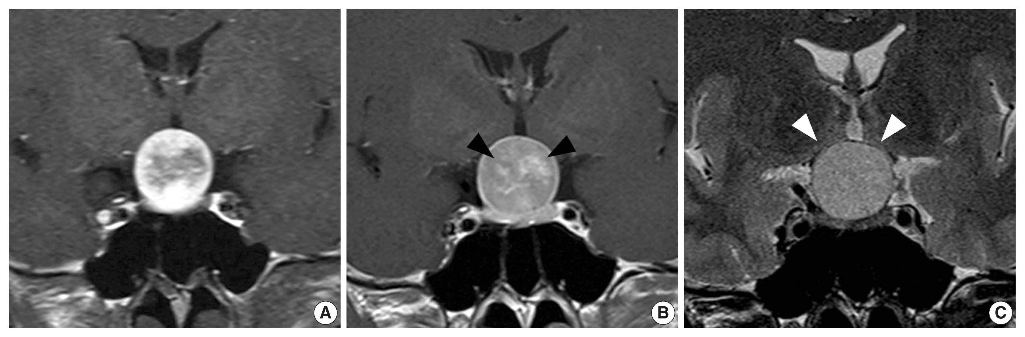
- Spindle cell oncocytoma of the sella turcica with anaplastic features and rapid progression in short-term follow-up: a case report with proposal of distinctive radiologic features
- Dong Ja Kim, SangHan Lee, Mee-seon Kim, Jeong-Hyun Hwang, Myong Hun Hahm
- J Pathol Transl Med. 2021;55(3):225-229. Published online March 9, 2021
- DOI: https://doi.org/10.4132/jptm.2021.01.27
- 4,358 View
- 107 Download
- 2 Web of Science
- 3 Crossref
-
Abstract
PDF
- We present a rare case of spindle cell oncocytoma (SCO) of the sella turcica with malignant histologic features and rapid progression. A 42-year-old woman experienced bilateral blurred vision and was preoperatively misdiagnosed as having a pituitary macroadenoma on magnetic resonance imaging. After surgery, SCO was diagnosed by the histopathologic features of interlacing fascicles of spindle tumor cells with finely granular, eosinophilic cytoplasm. Focal anaplastic changes and necrosis were present. Immunohistochemically, the tumor cells were positive for vimentin, epithelial membrane antigen, S-100, galectin-3, and thyroid transcription factor 1. Four months later, the tumor had progressed, and second surgery with adjuvant radiotherapy was performed; the patients remains under observation. In this report, we proposed distinctive radiologic features for differential diagnosis between SCO and other pituitary tumors.
-
Citations
Citations to this article as recorded by- Pituitary Spindle Cell Oncocytoma: More than a Grade 1 Tumor?
Jonathan Hammond, Zacharie Gagne, Bojana Mitrovic, Stefano M. Priola
Neurology International.2025; 17(2): 16. CrossRef - Treatment modalities and outcomes of granular cell tumors and spindle cell oncocytomas of the pituitary gland: an analysis of two national cancer databases
A. Yohan Alexander, Giorgos Michalopoulos, Panagiotis Kerezoudis, Jamie J. Van Gompel, Michael J. Link, Maria Peris-Celda
Acta Neurochirurgica.2024;[Epub] CrossRef - Spindle cell oncocytoma, a misdiagnosed rare entity of the pituitary – A case report with review of literature and special emphasis on the morphological differentials
Gittwa Vatsaraj Kottangal, Lilly Madhavan, Shalini Kuruvilla, Kavitha Kanjirakadu Parameswaran, Shehla Basheer Kollathodi
Indian Journal of Pathology and Oncology.2021; 8(4): 533. CrossRef
- Pituitary Spindle Cell Oncocytoma: More than a Grade 1 Tumor?
- Hepatoid thymic carcinoma: a case report of a rare subtype of thymic carcinoma
- Ji-Seon Jeong, Hyo Jeong Kang, Uiree Jo, Min Jeong Song, Soon Yeol Nam, Joon Seon Song
- J Pathol Transl Med. 2021;55(3):230-234. Published online April 14, 2021
- DOI: https://doi.org/10.4132/jptm.2021.03.10
- 4,121 View
- 120 Download
- 4 Web of Science
- 2 Crossref
-
Abstract
PDF
- Hepatoid thymic carcinoma is an extremely rare subtype of primary thymus tumor resembling “pure” hepatoid adenocarcinomas with hepatocyte paraffin 1 (Hep-Par-1) expression. A 53-year-old man presented with voice change and a neck mass. Multiple masses involving the thyroid, cervical and mediastinal lymph nodes, and lung were detected on computed tomography. Papillary thyroid carcinoma was confirmed by biopsy, and the patient underwent neoadjuvant chemoradiation therapy. However, the anterior mediastinal mass was enlarged after the treatment whereas the multiple masses in the thyroid and neck decreased in size. Microscopically, polygonal tumor cells formed solid sheets or trabeculae resembling hepatocytes and infiltrated remnant thymus. The tumor cells showed immunopositivity for cytokeratin 7, cytokeratin 19, and Hep-Par-1 and negativity for α-fetoprotein. Possibilities of germ cell tumor, squamous cell carcinoma, and metastasis of thyroid papillary carcinoma were excluded by immunohistochemistry. This report on the new subtype of thymic carcinoma is the third in English literature thus far.
-
Citations
Citations to this article as recorded by- Hepatoid thymic carcinoma in a polycythemia vera patient treated with ropeginterferon Alfa-2b: Clinical, histopathological and molecular correlates
Giuseppe G. Loscocco, Margherita Vannucchi, Raffaella Santi, Andrea Amorosi, Stefania Scarpino, Maria Chiara Siciliano, Paola Guglielmelli, Claudio Tripodo, Arianna Di Napoli, Alessandro M. Vannucchi
Pathology - Research and Practice.2024; 263: 155648. CrossRef - Hepatoid tumors of the gastrointestinal/pancreatobiliary district: morphology, immunohistochemistry, and molecular profiles
Paola Mattiolo, Aldo Scarpa, Claudio Luchini
Human Pathology.2023; 132: 169. CrossRef
- Hepatoid thymic carcinoma in a polycythemia vera patient treated with ropeginterferon Alfa-2b: Clinical, histopathological and molecular correlates
Editorials
- Histologic subtyping of ampullary carcinoma for targeted therapy
- Seung-Mo Hong
- J Pathol Transl Med. 2021;55(3):235-235. Published online May 13, 2021
- DOI: https://doi.org/10.4132/jptm.2021.04.28
- 3,045 View
- 117 Download
- 1 Web of Science
- Prognostic and predictive markers in glioblastoma and ALK overexpression
- Jang-Hee Kim
- J Pathol Transl Med. 2021;55(3):236-237. Published online May 13, 2021
- DOI: https://doi.org/10.4132/jptm.2021.04.29
- 3,941 View
- 94 Download
- 4 Web of Science
- 5 Crossref
-
PDF
-
Citations
Citations to this article as recorded by- Tumor markers in non-small cell lung cancer spine metastasis: an assessment of prognosis and overall survival
Brian Foresi, Aakash Shah, Seth Meade, Ajit Krishnaney
European Spine Journal.2024; 33(11): 4346. CrossRef - Extracellular Vesicle-Based Characterization of Stem Cell Phenotype in Glioblastomas
Georgiana M Serban, Manu Doina, Rodica Balasa, Adrian F Balasa
Cureus.2024;[Epub] CrossRef - CAR T-cells to treat brain tumors
Grace Guzman, Karolina Pellot, Megan R. Reed, Analiz Rodriguez
Brain Research Bulletin.2023; 196: 76. CrossRef - ALK fusions in the pan-cancer setting: another tumor-agnostic target?
Aditya Shreenivas, Filip Janku, Mohamed A. Gouda, Hui-Zi Chen, Ben George, Shumei Kato, Razelle Kurzrock
npj Precision Oncology.2023;[Epub] CrossRef - Glioblastoma multiforme targeted delivery of docetaxel using bevacizumab-modified nanostructured lipid carriers impair in vitro cell growth and in vivo tumor progression
Leonardo Delello Di Filippo, Jonatas Lobato Duarte, Juliana Hofstätter Azambuja, Rubia Isler Mancuso, Marcela Tavares Luiz, Victor Hugo Sousa Araújo, Ingrid Delbone Figueiredo, Lucas Barretto-de-Souza, Rafael Miguel Sábio, Estela Sasso-Cerri, Amanda Marti
International Journal of Pharmaceutics.2022; 618: 121682. CrossRef
- Tumor markers in non-small cell lung cancer spine metastasis: an assessment of prognosis and overall survival
 First
First Prev
Prev


