- Interobserver diagnostic reproducibility in advanced-stage endometrial carcinoma
-
Ho Jin Jung, Soo Yeon Lee, Jin Hwa Hong, Yi Kyeong Chun
-
J Pathol Transl Med. 2021;55(1):43-52. Published online December 3, 2020
-
DOI: https://doi.org/10.4132/jptm.2020.10.04
-
-
4,951
View
-
119
Download
-
5
Web of Science
-
4
Crossref
-
 Abstract Abstract
 PDF PDF
- Background
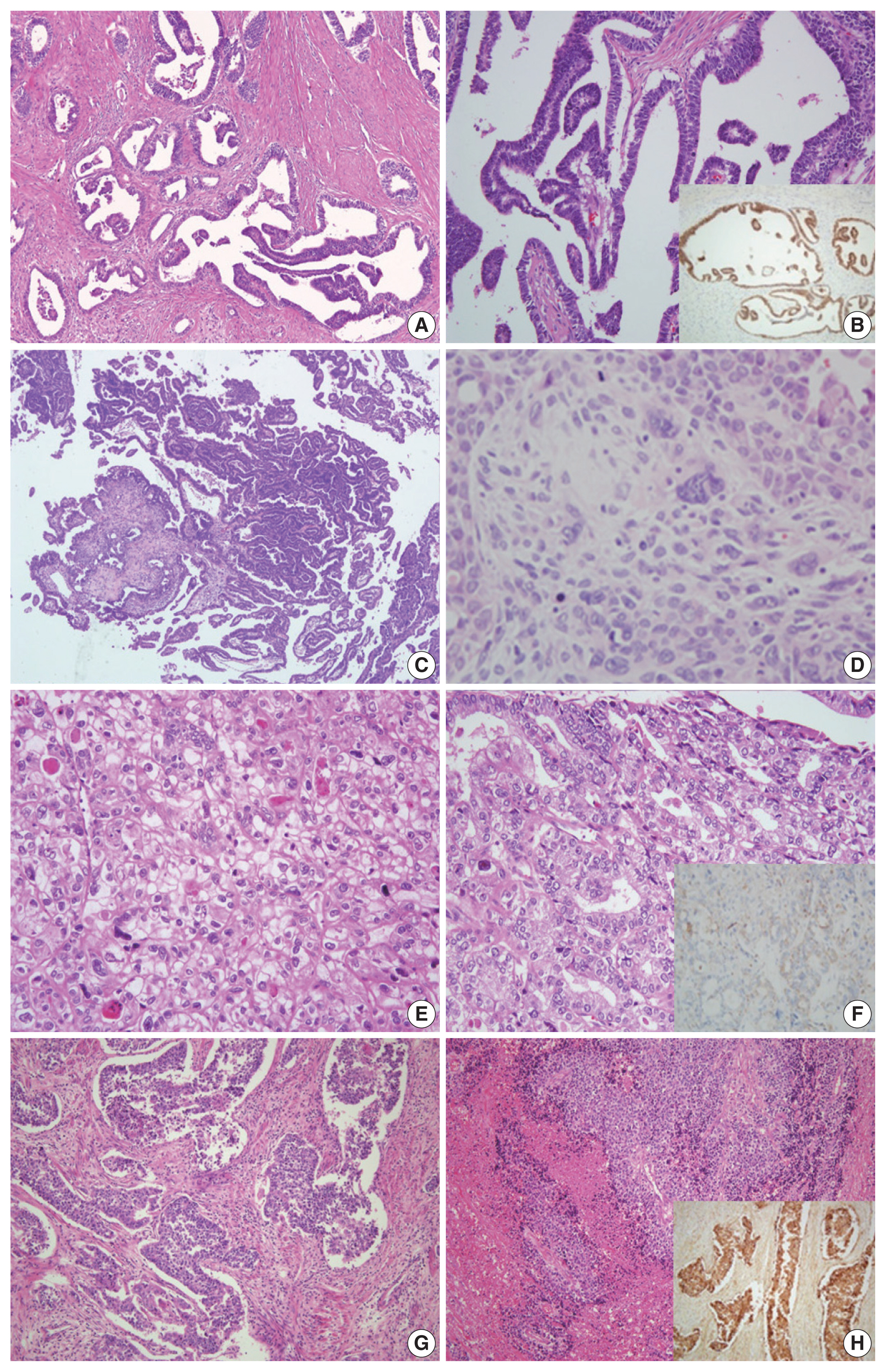
The accurate pathologic diagnosis and subtyping of high-grade endometrial carcinoma are often problematic, due to its atypical and overlapping histopathological features.
Methods
Three pathologists reviewed 21 surgically resected cases of advancedstage endometrial carcinoma. The primary diagnosis was based only on hematoxylin and eosin stained slides. When a discrepancy arose, a secondary diagnosis was made by additional review of immunohistochemical (IHC) stains. Finally, three pathologists discussed all cases and rendered a consensus diagnosis.
Results
The primary diagnoses were identical in 13/21 cases (62%). The secondary diagnosis based on the addition of IHC results was concordant in four of eight discrepant cases. Among four cases with discrepancies occurring in this step, two cases subsequently reached a consensus diagnosis after a thorough discussion between three reviewers. Next-generation sequencing (NGS) study was performed in two cases in which it was difficult to distinguish between serous carcinoma and endometrioid carcinoma. Based on the sequencing results, a final diagnosis of serous carcinoma was rendered. The overall kappa for concordance between the original and consensus diagnosis was 0.566 (moderate agreement).
Conclusions
We investigated stepwise changes in interobserver diagnostic reproducibility in advanced-stage endometrial carcinoma. We demonstrated the utility of IHC and NGS study results in the histopathological diagnosis of advanced-stage endometrial carcinoma.
-
Citations
Citations to this article as recorded by  - Accuracy of endometrial sampling in the diagnosis of endometrial cancer: a multicenter retrospective analysis of the JAGO-NOGGO
Zaher Alwafai, Maximilian Heinz Beck, Sepideh Fazeli, Kathleen Gürtler, Christine Kunz, Juliane Singhartinger, Dominika Trojnarska, Dario Zocholl, David Johannes Krankenberg, Jens-Uwe Blohmer, Jalid Sehouli, Klaus Pietzner
BMC Cancer.2024;[Epub] CrossRef - Deep Learning for Grading Endometrial Cancer
Manu Goyal, Laura J. Tafe, James X. Feng, Kristen E. Muller, Liesbeth Hondelink, Jessica L. Bentz, Saeed Hassanpour
The American Journal of Pathology.2024; 194(9): 1701. CrossRef - Application of NGS molecular classification in the diagnosis of endometrial carcinoma: A supplement to traditional pathological diagnosis
Qunxian Rao, Jianwei Liao, Yangyang Li, Xin Zhang, Guocai Xu, Changbin Zhu, Shengya Tian, Qiuhong Chen, Hui Zhou, Bingzhong Zhang
Cancer Medicine.2023; 12(5): 5409. CrossRef - Risk Stratification of Endometrial Cancer Patients: FIGO Stage, Biomarkers and Molecular Classification
Jenneke C. Kasius, Johanna M. A. Pijnenborg, Kristina Lindemann, David Forsse, Judith van Zwol, Gunnar B. Kristensen, Camilla Krakstad, Henrica M. J. Werner, Frédéric Amant
Cancers.2021; 13(22): 5848. CrossRef
- A Case of Malignant PEComa of the Uterus Associated with Intramural Leiomyoma and Endometrial Carcinoma
-
Yoo Jin Choi, Jin Hwa Hong, Aeree Kim, Hankyeom Kim, Hyeyoon Chang
-
J Pathol Transl Med. 2016;50(6):469-473. Published online July 25, 2016
-
DOI: https://doi.org/10.4132/jptm.2016.04.20
-
-
10,200
View
-
201
Download
-
8
Web of Science
-
8
Crossref
-
Abstract
PDF
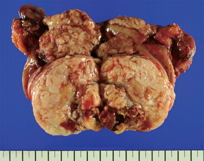
- Perivascular epithelioid cell tumors (PEComas) refers to a family of mesenchymal neoplasms composed of angiomyolipomas, clear cell “sugar” tumors of the lung, and lymphangioleiomyomatoses. These tumors have a distinctive and common component of perivascular epithelioid cells that show an association with blood vessel walls and immunohistochemically display myomelanocytic differentiation. The unique neoplasms have been shown to have an expanded range through a variety of case reports, including visceral, intra-abdominal, soft tissue, and bone tumors. The retroperitoneum, abdominopelvic region, and uterus have been reported to be the most common sites. Most PEComas follow a benign course. However, reports of malignant PEComas are increasing. Many papers have described uterine PEComas, but to our knowledge, there have not yet been any reports of a malignant PEComa arising concomitant with another epithelial tumor and mesenchymal tumor. We report herein the case of a 67-year-old woman who experienced a malignant uterine PEComa infiltrating a preexisting intramural leiomyoma with synchronous well differentiated endometrial carcinoma and multiple liver and lung metastases.
-
Citations
Citations to this article as recorded by - Risk prediction criteria for the primary hepatic perivascular epithelioid cell tumour family, including angiomyolipoma: analysis of 132 cases with a literature review
Youngeun Yoo, Jihun Kim, In Hye Song
Histopathology.2025; 86(6): 979. CrossRef - Metastasis of Clear Cell Renal Cell Carcinoma to Uterine Leiomyoma: First Case Report and Review of Literature
Sarvenaz Karamooz, Paula D. Binsol, Jaya Ruth Asirvatham, Anjali Pargaonkar
International Journal of Surgical Pathology.2024; 32(8): 1552. CrossRef - Uterine collision tumor (PEComa and endometrioid carcinoma) in a tuberous sclerosis patient: a case report
Nektarios Koufopoulos, Ioannis S. Pateras, Christos Koratzanis, Alina-Roxani Gouloumis, Argyro-Ioanna Ieronimaki, Alexandros Fotiou, Ioannis G. Panayiotides, Nikolaos Vrachnis
Frontiers in Oncology.2023;[Epub] CrossRef - TFE3-associated perivascular epithelioid cell tumor with complete response to mTOR inhibitor therapy: report of first case and literature review
Roli Purwar, Kishan Soni, Mridula Shukla, Ashish Verma, Tarun Kumar, Manoj Pandey
World Journal of Surgical Oncology.2022;[Epub] CrossRef - A case of perivascular epithelioid nodules arising in an intramural leiomyoma
Yoldez Houcine, Karima Mekni, Emna Brahem, Mouna Mlika, Aida Ayadi, Chiraz Fekih, Imene Ridene, Faouzi El Mezni
Human Pathology: Case Reports.2021; 23: 200470. CrossRef - Perivascular epithelioid cell tumors (PEComa) of the female genital tract: A challenging question for gynaecologic oncologist and pathologist
Angiolo Gadducci, Gian Franco Zannoni
Gynecologic Oncology Reports.2020; 33: 100603. CrossRef - Five cases of uterine perivascular epithelioid cell tumors (PEComas) and review of literature
Weiwei Shan, Yue Shi, Qin Zhu, Bingyi Yang, Liying Xie, Bing Li, Chengcheng Ning, Qiaoying Lv, Yali Cheng, Bingying Xie, Mingzhu Bai, Yuhui Xu, Xiaojun Chen, Xuezhen Luo
Archives of Gynecology and Obstetrics.2019; 299(1): 185. CrossRef - Uterine PEComas
Jennifer A. Bennett, Ana C. Braga, Andre Pinto, Koen Van de Vijver, Kristine Cornejo, Anna Pesci, Lei Zhang, Vicente Morales-Oyarvide, Takako Kiyokawa, Gian Franco Zannoni, Joseph Carlson, Tomas Slavik, Carmen Tornos, Cristina R. Antonescu, Esther Oliva
American Journal of Surgical Pathology.2018; 42(10): 1370. CrossRef
|
 E-submission