- Double cocktail immunostains with high molecular weight cytokeratin and GATA-3: useful stain to discriminate in situ involvement of prostatic ducts or acini from stromal invasion by urothelial carcinoma in the prostate
-
Junghye Lee, Youngeun Yoo, Sanghui Park, Min-Sun Cho, Sun Hee Sung, Jae Y. Ro
-
J Pathol Transl Med. 2020;54(2):146-153. Published online February 10, 2020
-
DOI: https://doi.org/10.4132/jptm.2019.11.12
-
-
6,812
View
-
126
Download
-
2
Web of Science
-
2
Crossref
-
 Abstract Abstract
 PDF PDF
- Background
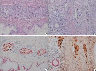
Distinguishing prostatic stromal invasion (PSI) by urothelial carcinoma (UC) from in situ UC involving prostatic ducts or acini with no stromal invasion (in situ involvement) may be challenging on hematoxylin and eosin stained sections. However, the distinction between them is important because cases with PSI show worse prognosis. This study was performed to assess the utility of double cocktail immunostains with high molecular weight cytokeratin (HMWCK) and GATA-3 to discriminate PSI by UC from in situ UC involvement of prostatic ducts or acini in the prostate.
Methods
Among 117 radical cystoprostatectomy specimens for bladder UCs, 25 cases showed secondary involvement of bladder UC in prostatic ducts/acini only or associated stromal invasion and of these 25 cases, seven cases revealed equivocal PSI. In these seven cases with equivocal PSI, HMWCK, and GATA-3 double immunohistochemical stains were performed to identify whether this cocktail stain is useful to identify the stromal invasion.
Results
In all cases, basal cells of prostate glands showed strong cytoplasmic staining for HMWCK and UC cells showed strong nuclear staining for GATA-3. In cases with stromal invasion of UC, GATA-3-positive tumor cells in the prostatic stroma without surrounding HMWCK-positive basal cells were highlighted and easily recognized. Among seven equivocal cases, two cases showed PSI and five in situ UC in the prostate. In two cases, the original diagnoses were revised.
Conclusions
Our study suggested that HMWCK and GATA-3 double stains could be utilized as an adjunct method in the distinction between PSI by UC from in situ UC involving prostatic ducts or acini.
-
Citations
Citations to this article as recorded by  - Aberrant expression of GATA3 in metastatic adenocarcinoma of the prostate: an important pitfall
João Lobo, Nazario P Tenace, Sofia Cañete‐Portillo, Isa Carneiro, Rui Henrique, Roberta Lucianò, Lara R Harik, Cristina Magi‐Galluzzi
Histopathology.2024; 84(3): 507. CrossRef - Utility of D2-40, Cytokeratin 5/6, and High–Molecular-weight Cytokeratin (Clone 34βE12) in Distinguishing Intraductal Spread of Urothelial Carcinoma From Prostatic Stromal Invasion
Oleksii A. Iakymenko, Laurence M. Briski, Katiana S. Delma, Merce Jorda, Oleksandr N. Kryvenko
American Journal of Surgical Pathology.2022; 46(4): 454. CrossRef
- Intraductal Carcinoma of Prostate: A Comprehensive and Concise Review
-
Jordan A. Roberts, Ming Zhou, Yong Wok Park, Jae Y. Ro
-
Korean J Pathol. 2013;47(4):307-315. Published online August 26, 2013
-
DOI: https://doi.org/10.4132/KoreanJPathol.2013.47.4.307
-
-
13,254
View
-
142
Download
-
11
Crossref
-
Abstract
PDF
Intraductal carcinoma of the prostate (IDC-P) is defined as a proliferation of prostate adenocarcinoma cells distending and spanning the lumen of pre-existing benign prostatic ducts and acini, with at least focal preservation of basal cells. Studies demonstrate that IDC-P is strongly associated with high-grade (Gleason grades 4/5), large-volume invasive prostate cancers. In addition, recent genetic studies indicate that IDC-P represents intraductal spread of invasive carcinoma, rather than a precursor lesion. Some of the architectural patterns in IDC-P exhibit architectural overlap with one of the main differential diagnoses, high-grade prostatic intraepithelial neoplasia (HGPIN). In these instances, additional diagnostic criteria for IDC-P, including marked nuclear pleomorphism, non-focal comedonecrosis (>1 duct showing comedonecrosis), markedly distended normal ducts/acini, positive nuclear staining for ERG, and cytoplasmic loss of PTEN by immunohistochemistry, can help make the distinction. This distinction between IDC-P and HGPIN is of critical importance because IDC-P has an almost constant association with invasive carcinoma and has negative clinical implications, including shorter relapse-free survival, early biochemical relapse, and metastatic failure rate after radiotherapy. Therefore, IDC-P should be reported in prostate biopsies and radical prostatectomies, regardless of the presence of an invasive component. This article will review the history, diagnostic criteria, molecular genetics, and clinical significance of IDC-P. -
Citations
Citations to this article as recorded by - Microfluidic Applications in Prostate Cancer Research
Kailie Szewczyk, Linan Jiang, Hunain Khawaja, Cindy K. Miranti, Yitshak Zohar
Micromachines.2024; 15(10): 1195. CrossRef - Detection limits of significant prostate cancer using multiparametric MR and digital rectal examination in men with low serum PSA: Up-date of the Italian Society of Integrated Diagnostic in Urology
Andrea B. Galosi, Erika Palagonia, Simone Scarcella, Alessia Cimadamore, Vito Lacetera, Rocco F. Delle Fave, Angelo Antezza, Lucio Dell'Atti
Archivio Italiano di Urologia e Andrologia.2021; 93(1): 92. CrossRef - Prostate cancer with comedonecrosis is frequently, but not exclusively, intraductal carcinoma: a need for reappraisal of grading criteria
Raghav Madan, Mustafa Deebajah, Shaheen Alanee, Nilesh S Gupta, Shannon Carskadon, Nallasivam Palanisamy, Sean R Williamson
Histopathology.2019; 74(7): 1081. CrossRef - The impact of intraductal carcinoma of the prostate on the site and timing of recurrence and cancer‐specific survival
Vincent Q. Trinh, Jennifer Sirois, Nazim Benzerdjeb, Babak K. Mansoori, Andrée‐Anne Grosset, Roula Albadine, Mathieu Latour, Anne‐Marie Mes‐Masson, Hélène Hovington, Alain Bergeron, Martin Ladouceur, Yves Fradet, Fred Saad, Dominique Trudel
The Prostate.2018; 78(10): 697. CrossRef - Comedonecrosis Revisited
Samson W. Fine, Hikmat A. Al-Ahmadie, Ying-Bei Chen, Anuradha Gopalan, Satish K. Tickoo, Victor E. Reuter
American Journal of Surgical Pathology.2018; 42(8): 1036. CrossRef - Focal Signet Ring Cell High-Grade Prostatic Intraepithelial Neoplasia on Needle Biopsy
Guang-Qian Xiao, Pamela D. Unger
International Journal of Surgical Pathology.2017; 25(4): 344. CrossRef - Exposure to maternal obesogenic diet worsens some but not all pre-cancer phenotypes in a murine genetic model of prostate cancer
Theresa Okeyo-Owuor, Emily Benesh, Scott Bibbey, Michaela Reid, Jacques Halabi, Siobhan Sutcliffe, Kelle Moley, Shree Ram Singh
PLOS ONE.2017; 12(5): e0175764. CrossRef - Histopathological features of intra-ductal carcinoma of prostatic and high grade prostatic intraepithelialneoplasia and correlation with PTEN and P63
Simin Torabi-Nezhad, Leila Malekmakan, Mohadese Mashayekhi, Arghavan Daneshian
The Prostate.2016; 76(4): 394. CrossRef - Intraduktales Karzinom der Prostata
G. Kristiansen, M. Varma, G. Seitz
Der Pathologe.2016; 37(1): 27. CrossRef - A Better Understating of the Morphological Features and Molecular Characteristics of Intraductal Carcinoma Helps Clinicians Further Explain Prostate Cancer Aggressiveness
Rodolfo Montironi, Liang Cheng, Antonio Lopez-Beltran, Marina Scarpelli, Francesco Montorsi
European Urology.2015; 67(3): 504. CrossRef - Clinicopathological analysis of intraductal proliferative lesions of prostate: intraductal carcinoma of prostate, high-grade prostatic intraepithelial neoplasia, and atypical cribriform lesion
Kosuke Miyai, Mukul K. Divatia, Steven S. Shen, Brian J. Miles, Alberto G. Ayala, Jae Y. Ro
Human Pathology.2014; 45(8): 1572. CrossRef
- Cellular Pseudosarcomatous Fibroepithelial Stromal Polyp of the Vagina during Pregnancy: A Lesion That Is Overdiagnosed as a Malignant Tumor
-
Joon Seon Song, Dong Eun Song, Kyu-Rae Kim, Jae Y. Ro
-
Korean J Pathol. 2012;46(5):494-498. Published online October 25, 2012
-
DOI: https://doi.org/10.4132/KoreanJPathol.2012.46.5.494
-
-
10,559
View
-
102
Download
-
9
Crossref
-
Abstract
PDF
Fibroepithelial stromal polyp (FSP) is a benign lesion that can occur at various sites, including the lower female genital tract. In rare cases, however, it may exhibit hypercellularity, bizarre cytomorphological features, and atypical mitoses resulting in an overdiagnosis as a malignant tumor despite its benign clinical course. Recently, we experienced one case of a 30-year-old pregnant woman with cellular pseudosarcomatous FSP that was initially diagnosed as a malignant fibrous histiocytoma at a primary clinic. In addition to describing the rare features of this case, we wish to increase awareness about this benign lesion which will be essential for avoiding unnecessary radical surgery or chemoradiation treatment. -
Citations
Citations to this article as recorded by - Giant hypopharyngeal fibroepithelial polyp: A case report and literature review
Muhammad Nour Alabdullah, Nagham Halaweek, Yasser Al Ghabra, Mohammad Hamdi, Mhd Ayham Abo Trab, Faysal Hajjar
Ear, Nose & Throat Journal.2025;[Epub] CrossRef - Fibroepithelial Polyp of the Vagina With Torsion: A Difficult Diagnosis Based on Clinical and Morphological Findings of the Vaginal Lesion
Efthymia Thanasa, Anna Thanasa, Gerasimos Kontogeorgis, Ektoras-Evangelos Gerokostas, Ioannis-Rafail Antoniou, Athanasios Chasiotis, Emmanouil M Xydias, Apostolos C Ziogas, Evangelos Kamaretsos, Ioannis Thanasas
Cureus.2024;[Epub] CrossRef - Recurrent fibroepithelial vaginal polyp in a two-year-old girl: A case report and review of the literature
Mohammad Hakam Shehadeh, Ahmad Abualrub, Waleed Malhes, Amar Msarweh, Wael Amro
Annals of Medicine & Surgery.2024;[Epub] CrossRef - A vaginal fibroepithelial stromal polyp: a case report with magnetic resonance images
Naoko Ogura, Mieko Inagaki, Ritsuko Yasuda, Shigeki Yoshida, Tetsuo Maeda
BJR|case reports.2022;[Epub] CrossRef - Fast-growing fibroepithelial stromal vaginal polyp
Ana Marta Pinto, Maria Boia Martins, Isabel Ferreira, Clara Moreira
BMJ Case Reports.2022; 15(6): e250076. CrossRef - Mesenchymal lesions of the vulva
David B. Chapel, Nicole A. Cipriani, Jennifer A. Bennett
Seminars in Diagnostic Pathology.2021; 38(1): 85. CrossRef - Giant Fibroepithelial Stromal Polyp of the Vulva: Diffusion-Weighted and Conventional Magnetic Resonance Imaging Features and Pathologic Correlation
Joonghyun Yoo, Bo-Kyung Je, Suk Keu Yeom, Ye Sul Park, Kyung-Jin Min, Joo Han Lee
Journal of Pediatric and Adolescent Gynecology.2019; 32(1): 93. CrossRef - Cellular Pseudosarcomatous Fibroepithelial Stromal Polyp of the Cervix: A Lesion Mimicking as Sarcoma
Ruquiya Afrose
Advances in Cytology & Pathology.2018;[Epub] CrossRef - Pseudosarcomatous Vaginal Polyp
Alexis Heller, Adanna Ukazu, Qing Wang
International Journal of Surgical Pathology.2017; 25(1): 54. CrossRef
|
 E-submission