 E-submission
Articles
- Page Path
- HOME > J Pathol Transl Med > Volume 48(4); 2014 > Article
-
Brief Case Report
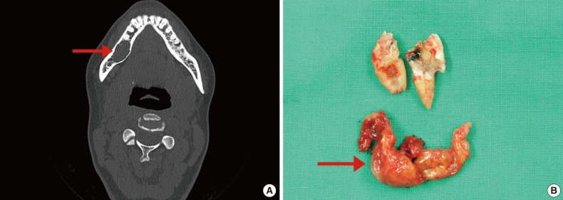
Solitary Myofibroma of the Adult Mandible: A Case Report and Review of Literature - Yong-Moon Lee, Seung-Myoung Son, Kyoung Won Kim1, Ok-Jun Lee
-
Korean Journal of Pathology 2014;48(4):307-310.
DOI: https://doi.org/10.4132/KoreanJPathol.2014.48.4.307
Published online: August 26, 2014
Department of Pathology, Chungbuk National University College of Medicine, Cheongju, Korea.
1Department of Oral and Maxillofacial Surgery, Chungbuk National University College of Medicine, Cheongju, Korea.
- Corresponding Author: Ok-Jun Lee, M.D. Department of Pathology, Chungbuk National University College of Medicine, 52 Naesudong-ro, Heungdeok-gu, Cheongju 361-763, Korea. Tel: +82-43-269-6260, Fax: +82-43-269-6269, 'ok5218@hanmail.net'
• Received: August 30, 2013 • Revised: September 26, 2013 • Accepted: September 27, 2013
© 2014 The Korean Society of Pathologists/The Korean Society for Cytopathology
This is an Open Access article distributed under the terms of the Creative Commons Attribution Non-Commercial License (http://creativecommons.org/licenses/by-nc/3.0/) which permits unrestricted non-commercial use, distribution, and reproduction in any medium, provided the original work is properly cited.

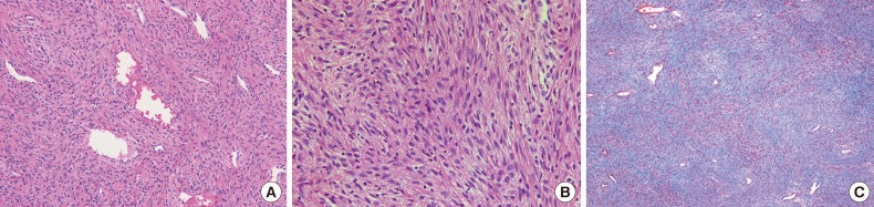
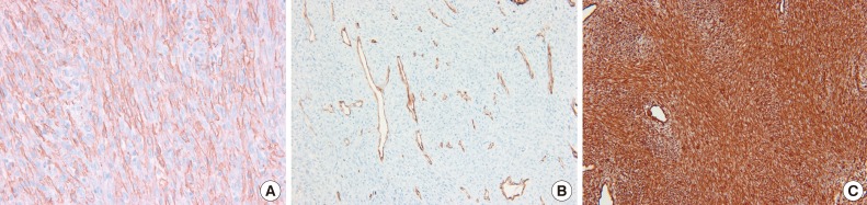
Figure & Data
References
Citations
Citations to this article as recorded by 
- Spontaneous resorption of an infantile mandibular myofibroma case report
Jason Azzi, Rajas Tipnis, Olivia Pieroni, Camelia Stefanovici, Darren J. Leitao
Acta Oto-Laryngologica Case Reports.2024; 9(1): 7. CrossRef - Myofibroma of the Gingiva – A Rare Case of Diagnostic Predicament
Arunima Sarma, Shraddha Jugade, Sunil Surendraprasad Mishra, Trupti Gaikwad, Shrutika Sonawane, Ashutosh S. Dighe
Journal of the International Clinical Dental Research Organization.2023; 15(2): 135. CrossRef - Intraosseous myofibroma of the mandible: A case report and review of the literature
Scott Cannon, Yousef Hammad, Thomas Schlieve
Oral and Maxillofacial Surgery Cases.2021; 7(4): 100234. CrossRef - Solitary intramuscular myofibroma in an adult: Case report and MR imaging findings
Irene Dixe de Oliveira Santo, Pedro V. Staziaki, Andrey Prilutskiy, Teviah E. Sachs, Akira M. Murakami
Clinical Imaging.2020; 67: 95. CrossRef - Infantile myofibromatosis treated by mandibulectomy and staged reconstruction with submental flap and free fibula flap: A case report
Alexandra Maby, Benoit Guay, François Thuot
Journal of Otolaryngology - Head & Neck Surgery.2019;[Epub] CrossRef - Adult Solitary Myofibroma of the Mandible Mimicking A Periapical Lesion
Jung-Hoon Yoon
The Korean Journal of Oral and Maxillofacial Pathology.2018; 42(5): 125. CrossRef - An update on myofibromas and myofibromatosis affecting the oral regions with report of 24 new cases
Molly Housley Smith, John D. Reith, Donald M. Cohen, Nadim M. Islam, Kimberly T. Sibille, Indraneel Bhattacharyya
Oral Surgery, Oral Medicine, Oral Pathology and Oral Radiology.2017; 124(1): 62. CrossRef - The spectrum of infantile myofibromatosis includes both non-penetrance and adult recurrence
Natalia Murray, B. Hanna, Nicole Graf, He Fu, Veronneau Mylène, P.M. Campeau, Anne Ronan
European Journal of Medical Genetics.2017; 60(7): 353. CrossRef - Solitary Intra-Osseous Myofibroma of the Jaw: A Case Report and Review of Literature
Anita Dhupar, Karla Carvalho, Poonam Sawant, Anita Spadigam, Shaheen Syed
Children.2017; 4(10): 91. CrossRef - Surgical Management of Myofibroma of the Gengiva
Paolo Garzino Demo, Matteo Savoini, Marco Marchetti, Francesca Maletta, Emanuele Zavattero, Guglielmo Ramieri
Journal of Craniofacial Surgery.2016; 27(7): e646. CrossRef
 PubReader
 ePub Link
-
 Cite this Article
- Cite this Article
-
- Close
- Download Citation
- Close
- Figure
-


- Related articles
-
- Primary leiomyosarcoma of the bone: a case report
- Metastatic choroidal melanoma in the breast: a case report and review of the literature
- Recurrent malignant solitary fibrous tumor of the scalp: a case report and literature review
- Renal intravascular large B cell lymphoma: the first case report in Korea and a review of the literature

