The evolving role of TRPS1 in dermatopathology: insights from the past 4 years
Article information

Abstract
Over the past 4 years, trichorhinophalangeal syndrome type 1 (TRPS1) has rapidly gained attention among practicing pathologists, with numerous studies emerging that both support and question its diagnostic utility. Initially regarded as a highly specific marker for tumors of mammary origin, TRPS1 is now recognized to have broader expression patterns, including in a variety of cutaneous neoplasms. This is likely due to embryologic parallels between breast tissue and skin adnexal structures, an overlap that was underappreciated in early investigations. Although TRPS1 lacks absolute specificity—even among cutaneous neoplasms—it can still offer meaningful diagnostic value when interpreted alongside conventional immunohistochemical markers and within the appropriate morphologic context. Noteworthy diagnostic applications include mammary Paget disease, primary extramammary Paget disease, rare adnexal neoplasms such as endocrine mucin-producing sweat gland carcinoma and primary cutaneous NUT adnexal carcinoma, and cutaneous metastases from breast carcinoma. In this review, we present the most comprehensive and up-to-date evaluation of the utility and limitations of TRPS1 immunohistochemistry in dermatopathology. Our aim is to deepen understanding of this emerging marker and provide practical guidance on its optimal integration with established immunohistochemical panels to enhance diagnostic accuracy in routine practice.
INTRODUCTION
Trichorhinophalangeal syndrome type 1 (TRPS1) immunohistochemistry (IHC) has gained significant traction among surgical pathologists and cytopathologists in recent years, initially being regarded as a highly sensitive and specific marker for carcinomas of mammary origin [1]. However, the study by Ai et al. [1], which first highlighted its potential, had a significant limitation: the authors employed tissue microarrays for analysis instead of whole tissue sections. This approach failed to account for the heterogeneity of TRPS1 expression within the same tumor types, undermining the robustness of their findings. Furthermore, their analysis excluded most cutaneous neoplasms, focusing primarily on melanomas, casting doubt on their assertion that TRPS1 expression is “highly specific” for breast carcinomas [1].
Subsequent studies have challenged this claim, demonstrating that TRPS1 expression extends beyond breast neoplasms. Tumors in sites with embryological similarities to breast tissue, such as skin adnexal structures, often share similar immunoprofiles (e.g., cytokeratin [CK] 7+/GATA3+). TRPS1 expression has been notably observed in various cutaneous adnexal neoplasms, including mammary Paget diseases (MPDs) [2,3], extramammary Paget diseases (EMPDs) [2-4], endocrine mucin-producing sweat gland carcinomas (EMPSGCs) [5], and numerous other cutaneous adnexal tumors [6-11].
In this review, we provide the most comprehensive and up-to-date examination of the utility and limitations of TRPS1 IHC in cutaneous neoplasms, encompassing not only adnexal tumors but also other epithelial and mesenchymal neoplasms of cutaneous origin. This knowledge will deepen our understanding of this emerging marker in dermatopathology and offer guidance on how to optimally integrate TRPS1 IHC with other established immunohistochemical markers to improve diagnostic accuracy in routine dermatopathology practice.
ANTI-TRPS1 ANTIBODIES
The successful detection of TRPS1 expression in formalin-fixed, paraffin-embedded (FFPE) tissues hinges on the selection of an optimal antibody and immunohistochemical protocol. Among the various anti-TRPS1 antibodies and clones reported in the literature, EPR16171, a rabbit monoclonal antibody, appears to be the most widely used for IHC [3,5-8,12]. However, its application varies across laboratories, with differences in antibody dilution (e.g., 1:2,000 [3,5,6,12] vs. 1:6,000 [7,8]), incubation times, antigen retrieval methods, and staining platforms.
The second most frequently employed anti-TRPS1 antibody is PA5-84587, a rabbit polyclonal antibody, used at diverse dilutions such as 1:100 [1], 1:250 [9], and 1:1,000 [4]. Another reported clone is ZR382, a rabbit monoclonal antibody used at a 1:200 dilution [13,14].
In our own experience, both monoclonal and polyclonal anti-TRPS1 antibodies have been evaluated. Notably, the monoclonal antibody EPR16171 reliably demonstrates TRPS1 expression in FFPE skin specimens. Internal controls, such as eccrine glands, consistently show strong (3+) expression intensity, supporting the reproducibility and robustness of this clone under our testing conditions.
TRPS1 EXPRESSION IN NORMAL SKIN
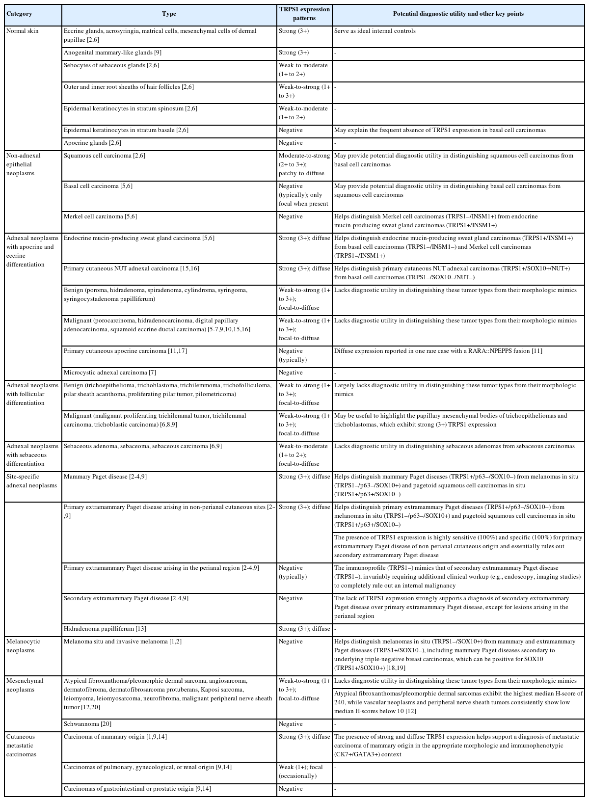
Understanding TRPS1 expression in the innate cellular and structural components of normal skin is crucial for identifying the types of cutaneous neoplasms that may express this marker. Neoplastic cells often retain the immunoprofile of their normal cellular counterparts as they proliferate and transform. In normal skin, TRPS1 immunoreactivity is prominently observed in various adnexal structures (Table 1), with the strongest expression (3+) found in eccrine glands (Fig. 1A), acrosyringia, matrical cells (Fig. 1B), and mesenchymal cells of the dermal papillae (Fig. 1C) [2,6]. These structures serve as ideal internal controls due to their consistent, robust TRPS1 expression. Sebocytes in sebaceous glands typically show weak-to-moderate (1+ to 2+) TRPS1 immunoreactivity, while the germinative cells of these glands are devoid of expression (Fig. 1D) [2,6]. Hair follicles exhibit variable TRPS1 expression in the outer and inner root sheaths and matrical cells, generally ranging from weak to strong intensity (1+ to 3+) (Fig. 1B) [2,6]. Arrector pili muscles also show natural TRPS1 immunoreactivity with weak-to-moderate intensity (1+ to 2+) (Fig. 1E). Notably, innate apocrine glands in normal skin do not express TRPS1 [2,6]. In contrast, anogenital mammary-like glands exhibit strong (3+) TRPS1 immunoreactivity [9].
Summary of TRPS1 immunoreactivity in normal skin and various cutaneous neoplasms
Trichorhinophalangeal syndrome type 1 (TRPS1) expression in normal skin. TRPS1 is strongly expressed in eccrine glands (A), matrical cells of hair follicles (B), and mesenchymal cells of the dermal papillae (C), making these structures reliable internal positive controls. Other skin adnexal structures, such as sebaceous glands (D) and arrector pili muscles (E), typically exhibit weak-to-moderate TRPS1 immunoreactivity (1+ to 2+). Notably, while sebocytes demonstrate TRPS1 expression, the germinative cells of sebaceous glands lack immunoreactivity (D, inset, arrows).
Regarding non-adnexal epithelial components, epidermal keratinocytes in the stratum spinosum may display weak-to-moderate (1+ to 2+) TRPS1 expression [2,6]. Although this TRPS1 immunoreactivity in innate epidermal keratinocytes was initially thought to be restricted to actinically damaged skin, it was later found that the epidermis from sun-protected sites can also occasionally express TRPS1 in weak-to-moderate (1+ to 2+) intensity [2,6]. The exact frequency of TRPS1 immunoreactivity in normal epidermal keratinocytes, as well as the comparison between sun-damaged and sun-protected skin, remains largely unknown at this point. Interestingly, while weak-to-moderate TRPS1 expression may be present in the normal epidermis, basal keratinocytes, unlike those in the stratum spinosum, naturally lack TRPS1 expression [2,6].
TRPS1 EXPRESSION IN CUTANEOUS NON-ADNEXAL EPITHELIAL NEOPLASMS
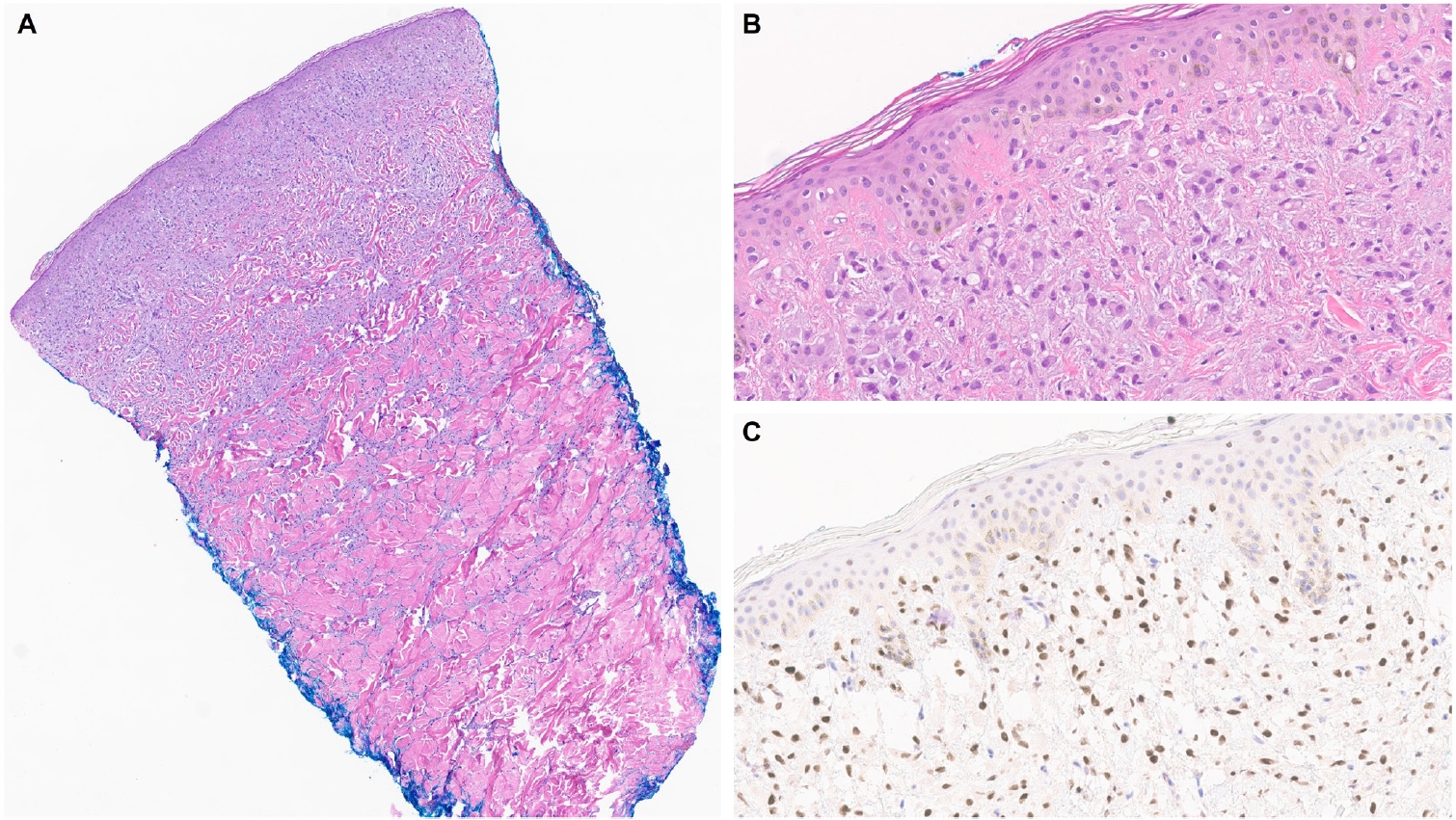
Most studies investigating TRPS1 expression in cutaneous non-adnexal epithelial neoplasms have focused on malignant tumors such as squamous cell carcinomas (SCCs), basal cell carcinomas (BCCs), and Merkel cell carcinomas (MCCs) (Table 1). A recent comprehensive study analyzing TRPS1 immunoreactivity across 200 cases of various cutaneous neoplasms found that nearly all SCCs (94%) demonstrated moderate to strong TRPS1 expression, with a median H-score of 200 [6]. Interestingly, the intensity of TRPS1 expression often decreases as SCC cells invade deeper into the dermis, whereas stronger expression tends to be retained in cells near the epidermis or within in situ components. This observation is supported by an earlier study showing diffuse and strong TRPS1 expression in 92% of SCC in situ (SCCIS) cases [2].
In contrast, the same study by Liu et al. [6] reported that the majority of BCCs (90%) either lacked TRPS1 expression entirely or showed only focal immunoreactivity, with a median H-score of 5. Given that BCCs are thought to originate from the basal layer of the interfollicular epidermis [21], the minimal TRPS1 expression may reflect the absence of TRPS1 expression in native basal keratinocytes. The difference in TRPS1 expression between SCCs and BCCs was statistically significant (p < .001) [6], highlighting its potential diagnostic utility in distinguishing between these entities. The authors further investigated whether squamous differentiation in BCCs might confound TRPS1-based discrimination. Even in BCCs with squamous differentiation, TRPS1 expression remained significantly more frequent in SCCs (p < .001) [6]. However, while these findings are statistically significant, clinical interpretation should be approached with caution, as exceptions do occur. For example, diffuse TRPS1 expression may occasionally be seen in BCCs with hamartomatous or infundibulocytic features and extensive squamous differentiation [6]. Conversely, TRPS1 expression may be nearly absent in rare cases of SCC [6,9].
Therefore, in diagnostically challenging scenarios, such as distinguishing BCCs with extensive squamous differentiation from SCCs with basaloid features, a comprehensive morphologic evaluation supplemented by an immunohistochemical panel, including TRPS1, epithelial membrane antigen, and BerEP4, may provide greater diagnostic accuracy. However, the utility of this proposed IHC panel in such cases warrants confirmation in larger, future studies.
MCCs consistently lack TRPS1 immunoreactivity, as demonstrated in prior studies [5,6]. This distinct immunophenotypic profile may be diagnostically useful in differentiating MCCs from EMPSGCs, a primary cutaneous adnexal carcinoma with neuroendocrine differentiation, which will be discussed in more detail below.
TRPS1 EXPRESSION IN CUTANEOUS ADNEXAL NEOPLASMS
Various benign and malignant cutaneous adnexal neoplasms have been shown to express TRPS1 with varying intensity and proportion [2-11,13,15,16,22], indicating that this marker is not specific to tumors of mammary origin (Table 1). However, TRPS1 generally lacks significant discriminatory power among most cutaneous adnexal neoplasms. Nonetheless, in certain diagnostic contexts, when used in combination with other immunohistochemical markers, TRPS1 may offer valuable diagnostic utility (Table 1).
TRPS1 EXPRESSION IN TUMORS WITH APOCRINE AND ECCRINE DIFFERENTIATION
Although not all tumor types that belong to this category of cutaneous adnexal neoplasms have been thoroughly investigated, published studies indicate that the majority, including poromas, hidradenomas (Fig. 2), spiradenomas, cylindromas, syringomas, mixed tumors, syringocystadenomas papilliferum, porocarcinomas, hidradenocarcinomas, digital papillary adenocarcinomas, squamoid eccrine ductal carcinomas, EMPSGCs, and primary cutaneous NUT adnexal carcinomas, express TRPS1 at least focally [5-7,9,10,15,16]. When present, TRPS1 expression in these tumors is typically weak-to-moderate (1+ to 2+), with the notable exception of EMPSGCs and primary cutaneous NUT adnexal carcinomas, which consistently demonstrate strong (3+) expression intensity [5,15,16], akin to that seen in eccrine glands, acrosyringia, or the mesenchymal cells of the dermal papillae. This consistent strong expression highlights the diagnostic utility of TRPS1 in identifying EMPSGCs and primary cutaneous NUT adnexal carcinomas.
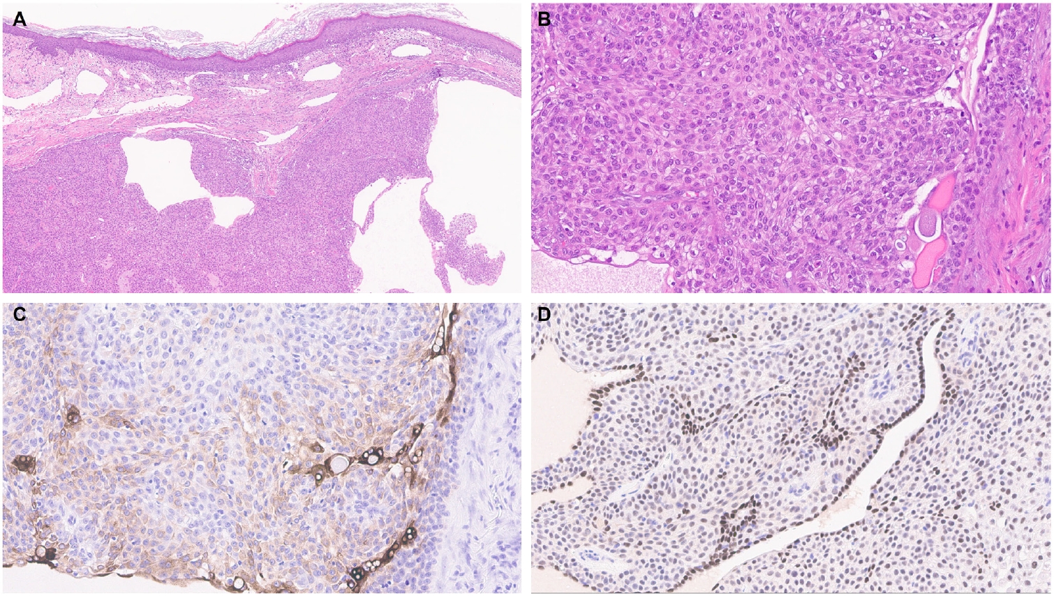
Hidradenoma. A biopsy from the left chest of a 62-year-old male patient reveals a solid and cystic hidradenoma (A, B). Cytokeratin 19 highlights ductal differentiation within the tumor (C). Intriguingly, while the majority of tumor cells show weak-to-moderate trichorhinophalangeal syndrome type 1 (TRPS1) immunoreactivity (1+ to 2+), the cuboidal cells lining the ductal structures exhibit strong TRPS1 expression (D).
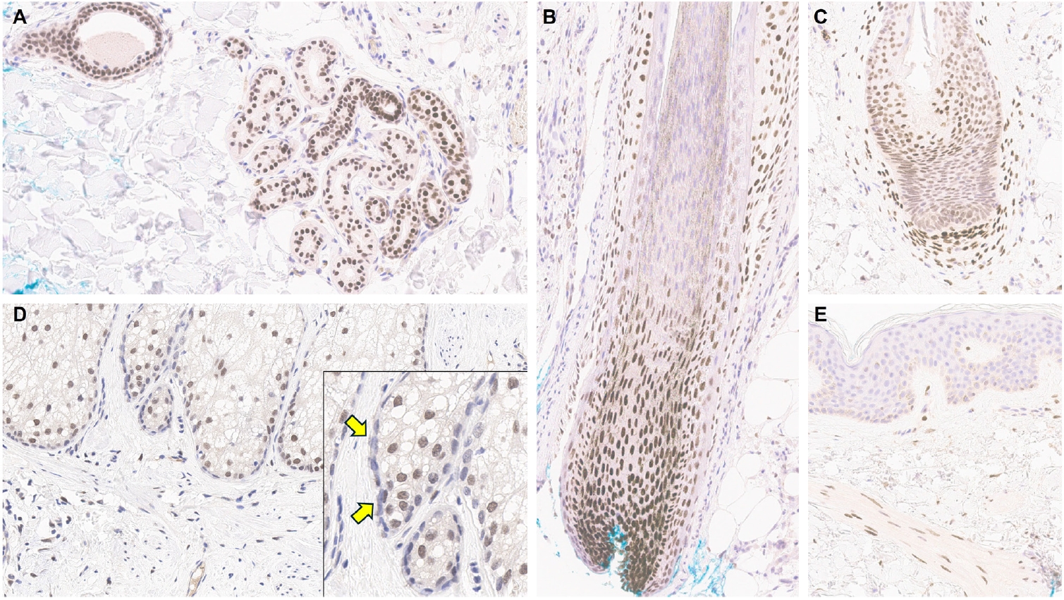
EMPSGCs are uncommon adnexal carcinomas of sweat gland origin with neuroendocrine differentiation and are often regarded as the cutaneous analogue of solid papillary carcinoma of the breast [23,24]. Histopathologically, EMPSGCs present as nodular proliferations of basaloid cells with varied architectural patterns and frequently contain intracellular and extracellular mucin, which can mimic BCCs, especially in superficial or shallow biopsies. Immunophenotypically, their expression of neuroendocrine markers such as insulinoma-associated protein 1 (INSM1), synaptophysin, and chromogranin may also lead to confusion with MCCs, another skin malignancy with neuroendocrine differentiation, albeit more common than EMPSGCs. A recent study evaluating TRPS1 expression in EMPSGCs, BCCs, and MCCs found that all EMPSGCs demonstrated diffuse, strong TRPS1 positivity, while most BCCs and all MCCs lacked TRPS1 immunoreactivity [5]. These findings support the role of TRPS1, particularly in combination with neuroendocrine markers such as INSM1, as a valuable immunohistochemical tool for differentiating EMPSGCs (Fig. 3) from their morphologic mimics, including BCCs and MCCs. Among neuroendocrine markers, INSM1 (Fig. 3D) is especially useful in this context due to its superior sensitivity compared to others like SOX11, synaptophysin, and chromogranin [25-27].
Endocrine mucin-producing sweat gland carcinoma. A biopsy from the right parietal scalp of a 71-year-old female patient demonstrates an endocrine mucin-producing sweat gland carcinoma (A, B). The tumor cells are diffusely positive for trichorhinophalangeal syndrome type 1 (TRPS1) (C) and insulinoma-associated protein 1 (D). In the absence of clinical or radiologic evidence of an underlying breast carcinoma, this immunoprofile in a cutaneous tumor is diagnostic of endocrine mucin-producing sweat gland carcinoma. Notably, TRPS1 expression in this tumor is consistently strong, comparable to that seen in native eccrine glands.
Primary cutaneous NUT adnexal carcinomas are a rare and recently characterized entity defined by recurrent NUTM1 or NUTM2B gene fusions [15,16,22,28]. These tumors typically exhibit a basaloid morphology, often resembling BCCs or other basaloid adnexal carcinomas such as porocarcinomas. Recent studies have shown that primary cutaneous NUT adnexal carcinomas consistently express TRPS1, SOX10, and NUT [15,16,28], suggesting that this unique immunoprofile can aid in distinguishing them from morphologically similar tumors such as porocarcinomas and BCCs. While porocarcinomas may express NUT in cases harboring a YAP1::NUTM1 fusion [29,30], they typically lack SOX10 expression [31], and TRPS1 expression in poroid neoplasms is generally not diffusely or strongly positive [6,7]. BCCs are typically negative for TRPS1, SOX10, and NUT, further supporting the diagnostic utility of this panel of IHC.
Among the primary cutaneous adnexal carcinomas studied to date, microcystic adnexal carcinomas were found to lack TRPS1 expression in one study [7]. Consistent with the observation that normal apocrine glands are also TRPS1-negative, primary cutaneous apocrine carcinomas were initially believed to be devoid of TRPS1 expression [17]. However, a recent case report described a primary cutaneous apocrine carcinoma harboring an RARA::NPEPPS fusion that showed diffuse TRPS1 positivity [11]. This finding underscores the need for additional studies with larger cohorts to clarify the true frequency and diagnostic relevance of TRPS1 expression in this rare tumor type.
TRPS1 EXPRESSION IN TUMORS WITH FOLLICULAR DIFFERENTIATION
Since hair follicles inherently express TRPS1, its presence in cutaneous adnexal neoplasms of hair follicle origin or those with follicular differentiation is expected rather than incidental. Several studies have documented TRPS1 expression, at least focally and with varying intensities, in tumors such as trichoepitheliomas, trichoblastomas, trichilemmomas, trichofolliculomas, pilar sheath acanthomas, proliferating pilar tumors, pilomatricomas, malignant proliferating trichilemmal tumors, trichilemmal carcinomas, and trichoblastic carcinomas [6,8,9].
While the diagnostic value of TRPS1 in these tumors remains somewhat limited, recent work suggests it may aid in differentiating trichoepitheliomas and trichoblastomas from BCCs, their morphologic mimics [8]. Mesenchymal cells in the dermal papillae typically exhibit strong (3+) TRPS1 expression, and this pattern is also seen in the papillary mesenchymal bodies of trichoepitheliomas and trichoblastomas. In contrast, BCCs, though they may show weak TRPS1 expression in peritumoral stromal cells, lack papillary mesenchymal bodies. While further systematic validation is needed, TRPS1 could offer additional diagnostic and discriminative value for dermatopathologists when used alongside other established markers, such as CK20 (for innate Merkel cells) and PHLDA1 (a follicular stem cell marker), especially in challenging cases (e.g., small biopsy samples) [32].
TRPS1 EXPRESSION IN TUMORS WITH SEBACEOUS DIFFERENTIATION
Sebaceous adenomas, sebaceomas, and sebaceous carcinomas typically show at least focal expression of TRPS1, with weak-to-moderate (1+ to 2+) intensities, reflecting the expression pattern seen in their cell of origin, sebocytes [6,9]. Due to the lack of specificity within this tumor category, TRPS1 offers limited diagnostic utility in distinguishing sebaceous adenomas from malignant counterparts like sebaceous carcinomas.
TRPS1 EXPRESSION IN SITE-SPECIFIC ADNEXAL TUMORS
One of the earliest and most promising applications of TRPS1 IHC in dermatopathology has been its supportive role in diagnosing MPDs and EMPDs [2-4,9]. This utility is grounded in the origin of Paget cells in MPDs, which are derived from the underlying ductal carcinoma of the breast. These cells exhibit strong epidermotropism within the nipple-areolar complex. Given that TRPS1 is highly expressed in nearly all breast carcinomas, including those with a triple-negative phenotype [1], it is not surprising that Paget cells in MPDs are also TRPS1-positive (Fig. 4). A seminal study confirmed this, demonstrating consistent TRPS1 expression in all MPD cases examined (100%; 24/24) [2].
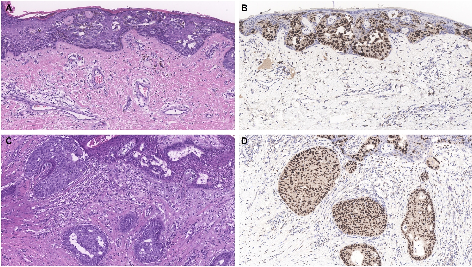
Mammary Paget disease. A biopsy of the left nipple from a 62-year-old woman demonstrates mammary Paget disease predominantly involving the nipple epidermis (A). Trichorhinophalangeal syndrome type 1 (TRPS1) shows strong, diffuse positivity within the intraepidermal Paget cells (B). Focal dermal involvement is also noted (C), corresponding to an invasive component that similarly exhibits strong and diffuse TRPS1 expression (D). Given that TRPS1 is highly expressed in nearly all breast carcinomas, and that mammary Paget disease is almost always secondary to an underlying breast carcinoma, TRPS1 serves as an ideal marker for confirming the diagnosis of mammary Paget disease in this context.
When evaluating a pagetoid intraepidermal neoplasm, such as MPD or EMPD, in the skin or nipple-areolar region, a panel of immunohistochemical studies is typically required to distinguish these entities from morphologic mimics, particularly pagetoid SCCIS and melanoma in situ (MIS). The reliable expression of TRPS1 in Paget cells is diagnostically helpful, particularly when interpreted alongside other markers such as CK7 (or Cam5.2), p63, and SOX10. For example, MPDs typically demonstrate immunoreactivity for CK7 (or Cam5.2) and TRPS1 (Fig. 4B), while lacking expression of p63 (or CK5/6) and SOX10 (or other melanocytic markers like Melan-A or HMB45). In contrast, SCCISs and MISs have distinct immunoprofiles. Although TRPS1 expression has been observed in SCCISs [2], this entity rarely involves the nipple-areolar complex. When SCCIS occurs at other anatomic sites, tumor cells are usually positive for p63 (or CK5/6) and negative for SOX10. Since low molecular weight cytokeratins such as CK7 and Cam5.2 can sometimes be expressed in SCCISs, especially those with prominent pagetoid features [33], their diagnostic utility in this context may be limited.
MISs, on the other hand, typically express SOX10 while lacking CK7 and p63 expression. Importantly, MISs are consistently negative for TRPS1 [2], making TRPS1 a particularly useful marker in challenging cases. This is especially relevant in MPD cases arising from underlying triple-negative or metaplastic breast carcinomas, which may express SOX10 [18,19]—a well-documented diagnostic pitfall. In such scenarios, the strong and diffuse expression of TRPS1 in intraepidermal pagetoid cells can support a diagnosis of MPD, even in the presence of SOX10 positivity.
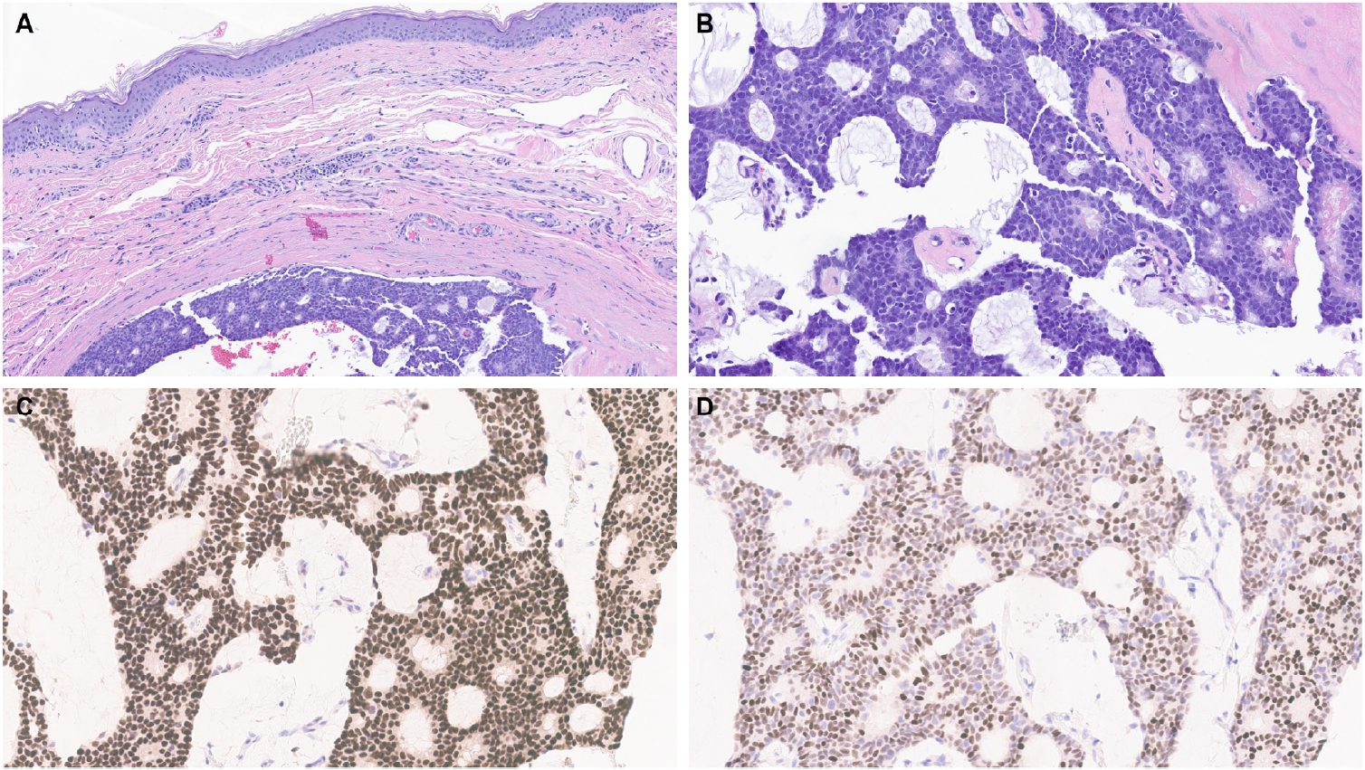
Similar to MPD, recent studies have shown that the majority of EMPD cases—ranging from 88% to 100%—also exhibit strong and diffuse TRPS1 expression [2-4,9]. Three key findings have emerged from these studies [2-4,9]: (1) TRPS1 is both highly sensitive (100%) and specific (100%) for primary EMPD arising outside the perianal region, such as the groin/inguinal area and axilla (Fig. 5); (2) secondary EMPDs, which originate from underlying internal malignancies such as colorectal or urothelial carcinomas, consistently lack TRPS1 expression (Fig. 6); and (3) the majority of perianal primary EMPDs—up to 91%—also lack TRPS1 expression, mimicking the immunoprofile of secondary EMPDs.
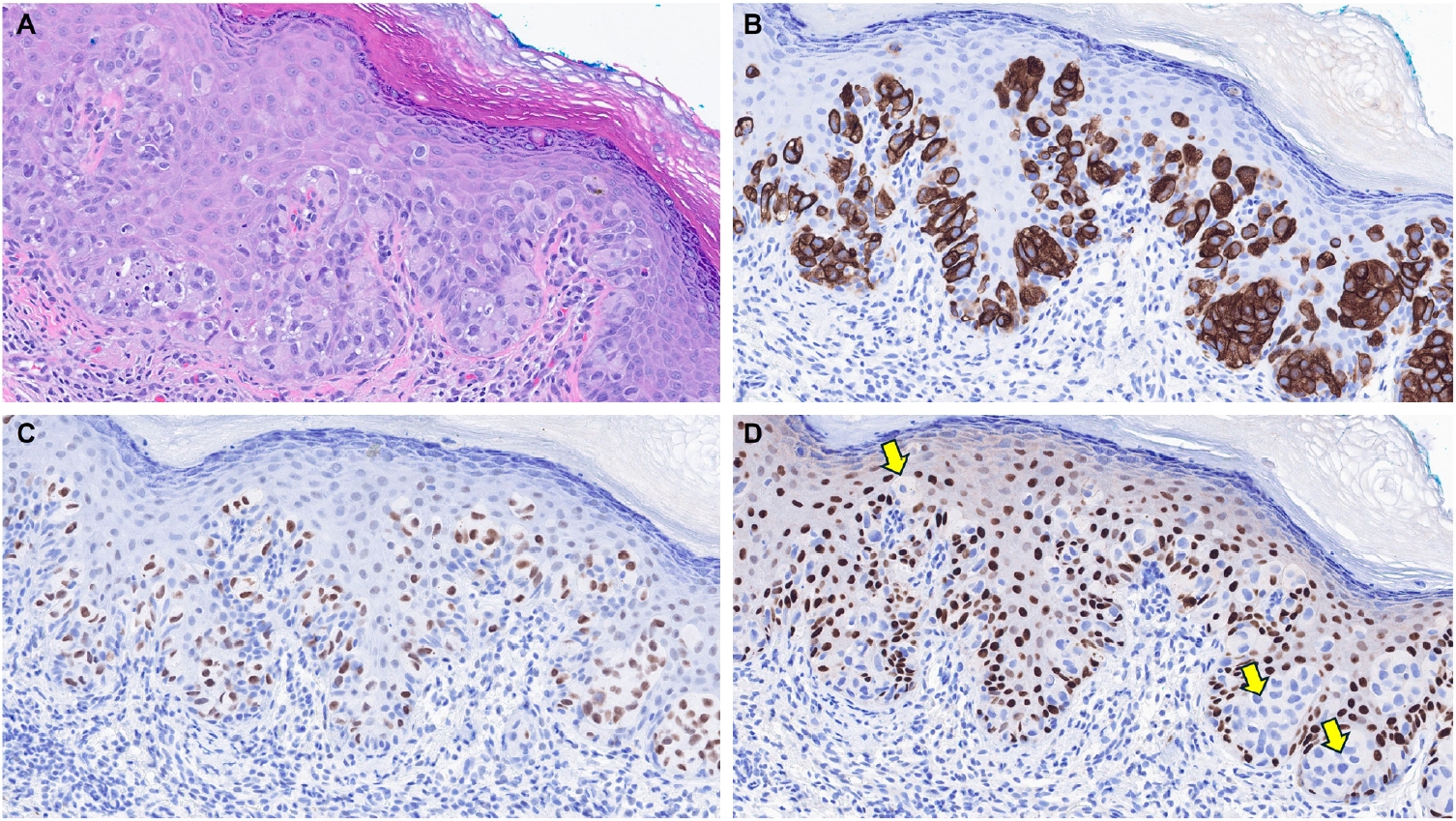
Primary extramammary Paget disease. A biopsy from the right medial thigh of a 63-year-old male patient reveals a pagetoid intraepidermal neoplasm (A). The lesional cells exhibit diffuse immunoreactivity for cytokeratin 7 (B) and trichorhinophalangeal syndrome type 1 (TRPS1) (C). In contrast, these cells show a complete absence of p63 immunoreactivity (D, arrows). Given that the neoplasm arises outside the perianal region, this immunoprofile is consistent with primary extramammary Paget disease.
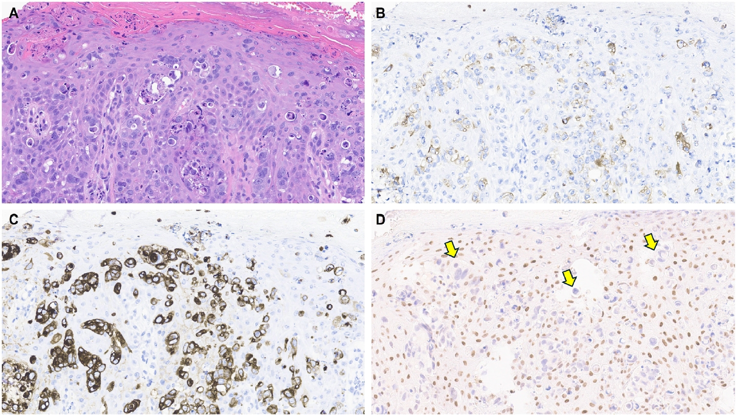
Secondary extramammary Paget disease. A biopsy from the perianal skin of a 79-year-old female patient with a known rectal adenocarcinoma shows a pagetoid intraepidermal neoplasm (A). The lesional cells exhibit patchy, weak positivity for cytokeratin (CK) 7 (B) and diffuse positivity for CK20 (C). These cells are completely negative for trichorhinophalangeal syndrome type 1 (TRPS1) (D, arrows). In the context of the underlying rectal adenocarcinoma, this immunoprofile is consistent with secondary extramammary Paget disease. Note that epidermal keratinocytes may occasionally be weakly immunoreactive for TRPS1 (D).
This third observation is especially diagnostically important. Given the high rate of TRPS1 negativity in perianal primary EMPDs, thorough clinical workup, including endoscopy or imaging, is essential to conclusively exclude an associated internal malignancy. Additional markers, such as CK20 and gross cystic disease fluid protein-15 (GCDFP-15), may aid in this distinction: a CK20-negative/GCDFP-15–positive immunoprofile may support a primary origin, while CK20 positivity with GCDFP-15 negativity leans toward a secondary source [34]. However, caution is warranted, as CK20 expression is not entirely specific—approximately one-third of perianal primary EMPDs can also express CK20 [34].
A few additional tumor types fall within this category, all of which are associated with anogenital mammary-like glands. These include hidradenoma papilliferum (HP), fibroadenoma and phyllodes tumor of anogenital mammary-like glands, and adenocarcinoma of mammary gland type. HPs, believed to originate from anogenital mammary-like glands, most commonly occur in the vulva and only rarely in the perianal region. Given that native anogenital mammary-like glands demonstrate TRPS1 expression [9], it is not surprising that a recent study found consistent TRPS1 positivity in all HPs (100%; 9/9), except within intratumoral foci exhibiting oxyphilic metaplasia, which lacked expression [13].
Due to the rarity of other neoplasms in this group, data on TRPS1 expression in tumors such as adenocarcinomas of mammary gland type remain limited. However, based on the authors’ limited experience, TRPS1 immunoreactivity has been observed in at least one case of adenocarcinoma of mammary gland type, although larger studies are needed to confirm and better characterize this finding.
TRPS1 EXPRESSION IN MELANOCYTIC NEOPLASMS
MISs and invasive melanomas are essentially devoid of TRPS1 expression (Table 1) [1,2]. While benign melanocytic neoplasms such as common (banal) nevi have not been well studied with regard to TRPS1 immunoreactivity, they are also likely negative for TRPS1, given that normal epidermal melanocytes do not express this marker. However, the TRPS1 expression profile in other melanocytic neoplasms, including melanocytomas such as WNT-activated deep penetrating/plexiform melanocytomas and pigmented epithelioid melanocytomas, remains largely uncharacterized.
TRPS1 EXPRESSION IN CUTANEOUS MESENCHYMAL NEOPLASMS
Limited studies have explored TRPS1 immunoreactivity in non-epithelial neoplasms (Table 1). One recent study assessed TRPS1 expression in 135 cases of various cutaneous mesenchymal neoplasms and tumors of uncertain differentiation, including atypical fibroxanthomas (AFXs) (Fig. 7A, B). TRPS1 was frequently expressed in dermatofibromas (100%; 24/24), leiomyomas (100%; 8/8), AFXs/pleomorphic dermal sarcomas (PDSs) (95%; 20/21), leiomyosarcomas (75%; 6/8), and dermatofibrosarcomas protuberans (64%; 14/22) (Fig. 7C, D) [12]. Expression was less frequent in neurofibromas (29%; 5/17), Kaposi sarcomas (25%; 2/8), and angiosarcomas (15%; 3/20), and was completely absent in perineuriomas [12]. Considering both the proportion and intensity of TRPS1 expression, AFXs/PDSs exhibited the highest median H-score of 240, whereas vascular neoplasms and peripheral nerve sheath tumors had consistently low median H-scores below 10 [12].
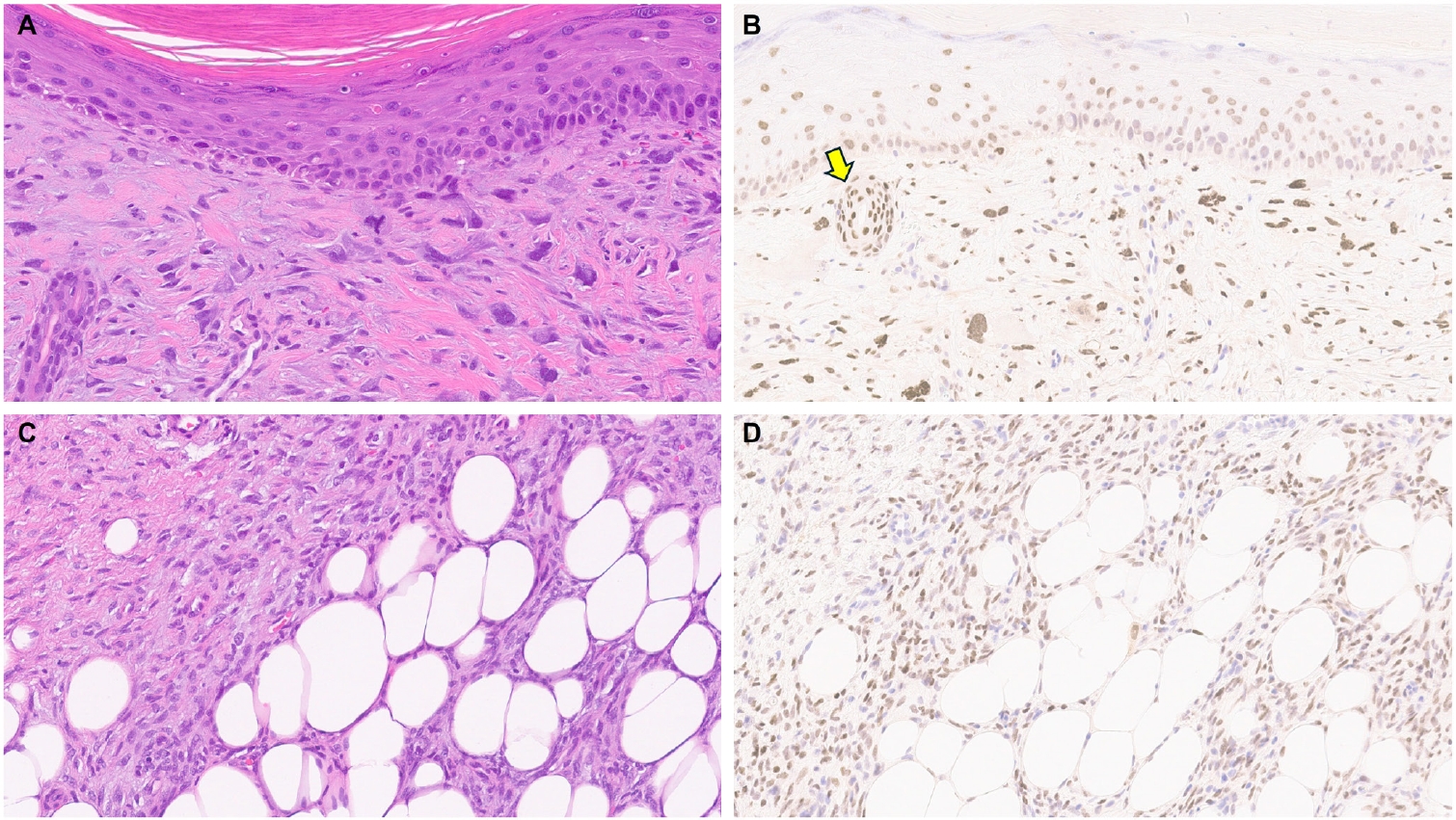
Atypical fibroxanthoma and dermatofibrosarcoma protuberans. A biopsy from the left frontal scalp of a 72-year-old male patient reveals a dermal proliferation of pleomorphic spindled and epithelioid cells that are negative for cytokeratin cocktail, p40, SOX10, ERG, and desmin, but positive for CD10, consistent with atypical fibroxanthoma (A). The tumor cells exhibit diffuse and strong positivity for trichorhinophalangeal syndrome type 1 (TRPS1) (B). Notably, the intensity of TRPS1 expression in these atypical fibroxanthoma cells is similar to that in eccrine sweat ducts (arrow), which serve as an internal control known to show 3+ TRPS1 immunoreactivity. A radical resection from the left groin of a 42-year-old male patient shows dermatofibrosarcoma protuberans (C), which may occasionally demonstrate TRPS1 expression (D).
The morphologic differential diagnosis of AFX often includes poorly differentiated or sarcomatoid SCC, melanoma, angiosarcoma, and leiomyosarcoma. The aforementioned study also examined the potential of TRPS1 in distinguishing AFXs from these morphologic mimics. While significant differences in H-scores were observed between AFXs and angiosarcomas (p < .001), melanomas (p < .001), and leiomyosarcomas (p = .029), no significant difference was noted when compared to sarcomatoid SCCs, suggesting limited discriminatory value of TRPS1 in this context [12].
Another recent study highlighted that a subset of malignant peripheral nerve sheath tumors also expresses TRPS1, albeit generally with weak intensity [20]. In contrast, other benign peripheral nerve sheath tumors, such as schwannomas and neurofibromas, consistently lacked TRPS1 expression [20].
Together, these studies provide new insights into TRPS1 expression patterns in a subset of cutaneous mesenchymal neoplasms and tumors of uncertain differentiation. This extends beyond prior research primarily focused on epithelial tumors and underscores potential limitations associated with TRPS1 IHC.
Finally, TRPS1 expression is not confined to neoplastic processes in the skin. Similar to its expression in cutaneous mesenchymal neoplasms of fibroblastic, myofibroblastic, or fibrohistiocytic origin, TRPS1 is also found in non-neoplastic stromal cells like fibroblasts and myofibroblasts, particularly in wound healing and scar formation [35]. This may be linked to TRPS1’s role as a key regulator in tissue regeneration. Notably, down-regulation of TRPS1 has been shown to promote epithelial-mesenchymal transition (EMT) and metastasis in various cancer types by repressing FOXA1, a negative regulator of EMT [36].
TRPS1 EXPRESSION IN CUTANEOUS METASTATIC CARCINOMAS
Given the well-documented high frequency of TRPS1 expression in breast carcinomas [1], it is not surprising that 95%–100% of metastatic mammary carcinomas involving the skin demonstrate strong and diffuse TRPS1 immunoreactivity (Table 1) [9,14]. In contrast, only focal and weak TRPS1 immunoreactivity may occasionally be observed in metastatic carcinomas of pulmonary (particularly SCCs), gynecologic, or renal origin (Table 1) [9,14]. Notably, carcinomas originating from the gastrointestinal tract, such as colonic and gastric adenocarcinomas, or from the prostate, typically lack TRPS1 expression (Table 1) [9,14]. Therefore, in the appropriate morphologic and immunophenotypic context (e.g., CK7+/GATA3+), strong and diffuse TRPS1 expression in a suspected case of cutaneous metastasis can support a mammary origin (Fig. 8).
Cutaneous metastasis of breast lobular carcinoma. A biopsy from the right abdomen of a 68-year-old female patient with a history of mammary lobular carcinoma reveals an infiltrating carcinoma involving the dermis, characterized by prominent histiocytoid morphology (A, B). The tumor cells show strong and diffuse nuclear positivity for trichorhinophalangeal syndrome type 1 (TRPS1) (C), supporting the diagnosis of cutaneous metastasis from the patient’s known breast carcinoma.
However, a key diagnostic limitation is that TRPS1 expression does not distinguish between a primary cutaneous adnexal carcinoma, including primary cutaneous apocrine carcinoma, and a cutaneous metastasis of breast carcinoma, as both can exhibit strong and diffuse TRPS1 positivity. In such cases, thorough clinicopathologic correlation, including imaging studies, is essential for accurate classification.
CONCLUSION
It is well recognized that the initially reported sensitivity and specificity of a new immunohistochemical marker often decline over time as its use becomes more widespread and as additional data and real-world experiences accumulate. TRPS1 IHC is no exception. Initially regarded as a highly sensitive and specific marker for breast carcinomas [1], TRPS1 has since been shown to be expressed in a broader range of cutaneous neoplasms, challenging its specificity.
As demonstrated throughout this review, TRPS1 expression is not exclusive to any single cutaneous tumor type. Nevertheless, when interpreted in conjunction with conventional markers and within the proper morphologic and immunophenotypic context, TRPS1 can offer valuable diagnostic insight. Notable examples include its use in diagnosing MPD (TRPS1+/CK7+/SOX10-/p63–), primary EMPD (TRPS1+/CK7+/SOX10–/p63–), primary cutaneous NUT adnexal carcinoma (TRPS1+/SOX10+/NUT+), EMPSGC (TRPS1+/INSM1+), and cutaneous metastases of mammary carcinoma (TRPS1+/CK7+/GATA3+). Although immunohistochemical studies are typically not necessary to differentiate SCCs from BCCs, the stark contrast in TRPS1 immunoreactivity (TRPS1+ in SCCs and TRPS1– in BCCs) may possibly provide additional diagnostic value.
We hope this review has provided a comprehensive and practical overview of the diagnostic utility and limitations of TRPS1 in dermatopathology. Ultimately, it is critical to emphasize that immunohistochemical findings must always be interpreted in the context of the overall morphologic features, and no single marker, regardless of its perceived sensitivity or specificity, should be used in isolation.
Notes
Ethics Statement
All procedures performed in the current review were approved by the IRB of the University of Texas MD Anderson Cancer Center (IRB#: 2022-0662) in accordance with the 1964 Helsinki declaration and its later amendments. Formal written informed consent was not required with a waiver by the appropriate IRB.
Availability of Data and Material
The data of this review are available from the corresponding author on reasonable request.
Code Availability
Not applicable.
Author Contributions
Conceptualization: WCC. Data curation: MHA. Formal analysis: MHA. Funding acquisition: WCC. Investigation: all authors. Methodology: WCC. Project administration: WCC. Resources: WCC. Software: N/A. Supervision: WCC. Validation: all authors. Visualization: all authors. Writing—original draft preparation: all authors. Writing—review & editing: all authors. Approval of final manuscript: all authors.
Conflicts of Interest
The authors declare that they have no potential conflicts of interest.
Funding Statement
This review was supported in part by the Institutional Start-up Funds from the University of Texas MD Anderson Cancer Center awarded to WCC.