Breast schwannoma: review of entity and differential diagnosis
Article information

Abstract
Schwannomas are benign peripheral nerve sheath tumors composed of Schwann cells, which uncommonly involve the breast. Most breast schwannomas are clinically present as a superficial palpable breast mass but may also be detected on screening mammography. Excision is the preferred treatment if symptomatic, and these are not known to recur. Histomorphology is similar to other anatomic sites: bland spindle cells with wavy nuclei, nuclear palisading (Verocay bodies), variably hypercellular (Antoni A) and hypocellular (Antoni B) areas, myxoid stroma, hyalinized vessels and variable cystic degeneration. Classic immunohistochemistry is diffuse and strong labeling for S100 and Sox10. Notable diagnostic pitfalls specific to the breast include myofibroblastoma, particularly the palisaded variant, and fascicular pseudoangiomatous stromal hyperplasia.
INTRODUCTION
Schwannomas are a well-described entity in soft tissue. These are benign peripheral nerve sheath tumors, composed of Schwann cells, most commonly occur in the head, trunk, and flexor surfaces of upper and lower extremities. Less described due to their rare nature are schwannomas that occur in breast parenchyma (2.6%) or axilla (5%) [1,2]. Although they resemble those of other anatomic sites, they pose a diagnostic pitfall with other spindle cell tumors in the breast [3]. The accurate diagnosis of this neoplasm, especially on small core biopsy specimens, plays a critical role for appropriate patient management and therapeutic options.
EPIDEMIOLOGY AND PATHOGENESIS
Schwannomas most often occur in adults in the third to fifth decade of life, with equal occurrences in males and females [1]. The pathogenesis of schwannomas is not well understood, although there are several hypotheses. One hypothesis is a genetic mutation or sporadic change leads to loss of merlin, which then causes overexpression of growth factors. This leads to tumorigenesis with decreased cell cycle arrest [4]. Another hypothesis involves genetic mutation(s) causing peripheral nerves to be vulnerable to stress and injury, which leads to unregulated Schwann cell proliferation and tumorigenesis [5].
MOLECULAR FINDINGS
Most schwannomas in the breast and other anatomic sites occur sporadically (90%). The remainder of schwannomas are attributed to various genetic alterations, and multiple occurrences may also be syndromic. A well described syndrome is Carney’s complex, which harbors PRKAR1a mutations [6], psammomatous melanotic schwannomas predominantly in the upper gastrointestinal tract and sympathetic chain, myxomas and Sertoli cell tumors [4]. Neurofibromin 2 (NF2)–related schwannomatosis due to alterations in NF2 [7] presents with numerous peripheral nerve sheath tumors, unilateral vestibular schwannoma, and meningioma [6]. Other types of schwannomatosis are due to loss of heterozygosity of chromosome 22q [7], and missense and truncating mutations of DGCR8 [6], LZTR1 [6], and SMARCB1 [6].
CLINICAL FEATURES AND RADIOLOGY FINDINGS
Majority of breast schwannomas present as a palpable, mobile, and nontender mass [8], although pain has been reported in some cases [9]. Breast schwannomas may also be incidentally detected on screening mammography. Using the Breast Imaging Reporting and Data System (BI-RADS), a system for standardizing mammogram reporting, these are often reported as 4A [8]. This category is considered suspicious for malignancy and is followed by an ultrasound and core biopsy [10].
On mammography, schwannomas present as a well-circumscribed, oval shaped, hyperdense nodule without microcalcifications (Fig. 1A). Ultrasound demonstrates a round/oval, well-circumscribed, homogenous, hypoechoic nodule with parallel orientation (Fig. 1B) [11,12]. Although not routinely performed, magnetic resonance imaging (MRI) is another modality to examine breast schwannomas. MRI T1 demonstrates a low signal and isointense nodule, while MRI T2 demonstrates a heterogeneous hyperintense signal with strong homogeneous contrast enhancement [13].
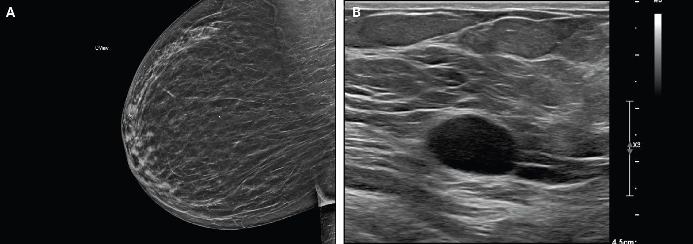
Mammogram with a well-circumscribed oval mass with equal density. The arrow indicates the lesion (A). Ultrasound with an oval hypoechoic mass with well-circumscribed margins (B).
PROGNOSIS AND TREATMENT
Breast schwannomas are benign tumors with a favorable prognosis. Expectant management is appropriate if the lesion is stable and patient is asymptomatic. Surgical excision is recommended in cases where the lesion is growing or patient is symptomatic. There is no evidence of recurrence after excision [14].
PATHOLOGIC FINDINGS
Gross description
Schwannomas have a similar gross appearance across anatomic locations, and none are unique to the breast. These often have a tan or yellow cut surface and are well demarcated from adjacent breast stroma. Degrees of hemorrhage and cystic changes are variable [4,15]. The plexiform variant of schwannoma has a distinct multinodular architecture [4].
Frozen sections
Features on frozen section for schwannomas include a bland spindle cell proliferation with various degrees of cellularity (Fig. 2) [16,17]. Individual cells demonstrate anisonucleosis, and have wavy, elongated nuclei with tapering ends. These are arranged in parallel along the fibrillary and variably collagenous stroma. Hemosiderin deposition may also be present [17]. It is important to note that frozen artifact is often prominent, which includes cytoplasmic vacuolization and gaps between collagen. Nuclear freezing artifact may also lead to an incorrect impression of malignancy [18].
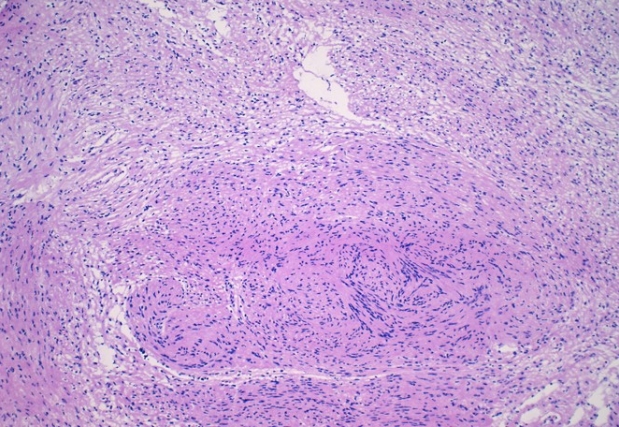
Intraoperative frozen section of schwannoma demonstrating a spindle cell proliferation with Verocay bodies, and variable cellularity (Antoni A and B). Clear cell change and white gaps in tissue are due to frozen section artifact.
Histomorphology
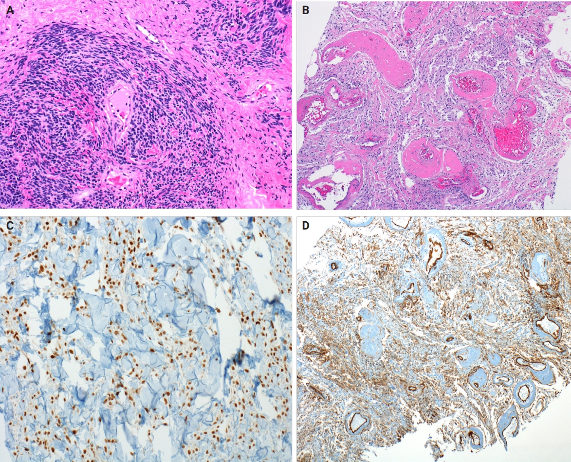
Microscopically, breast schwannomas are well-circumscribed or encapsulated and can have prominent nodularity (Fig. 3A). Classic schwannomas have a bland spindle cell proliferation with various degrees of anisonucleosis, and wavy, elongated nuclei with tapering ends. These are arranged in parallel rows (nuclear palisading), also known as Verocay bodies (Fig. 3B). There is an abrupt transition between hypercellular (Antoni A) and hypocellular areas (Antoni B) (Fig. 3C). Antoni B areas have loose and myxoid stroma (Fig. 4A). Other key features to schwannomas include numerous small to medium sized vessels with prominent hyalinization and thrombi inside the lumen (Fig. 4B) and may also contain areas of hemorrhage or hemosiderin deposition [3,19].
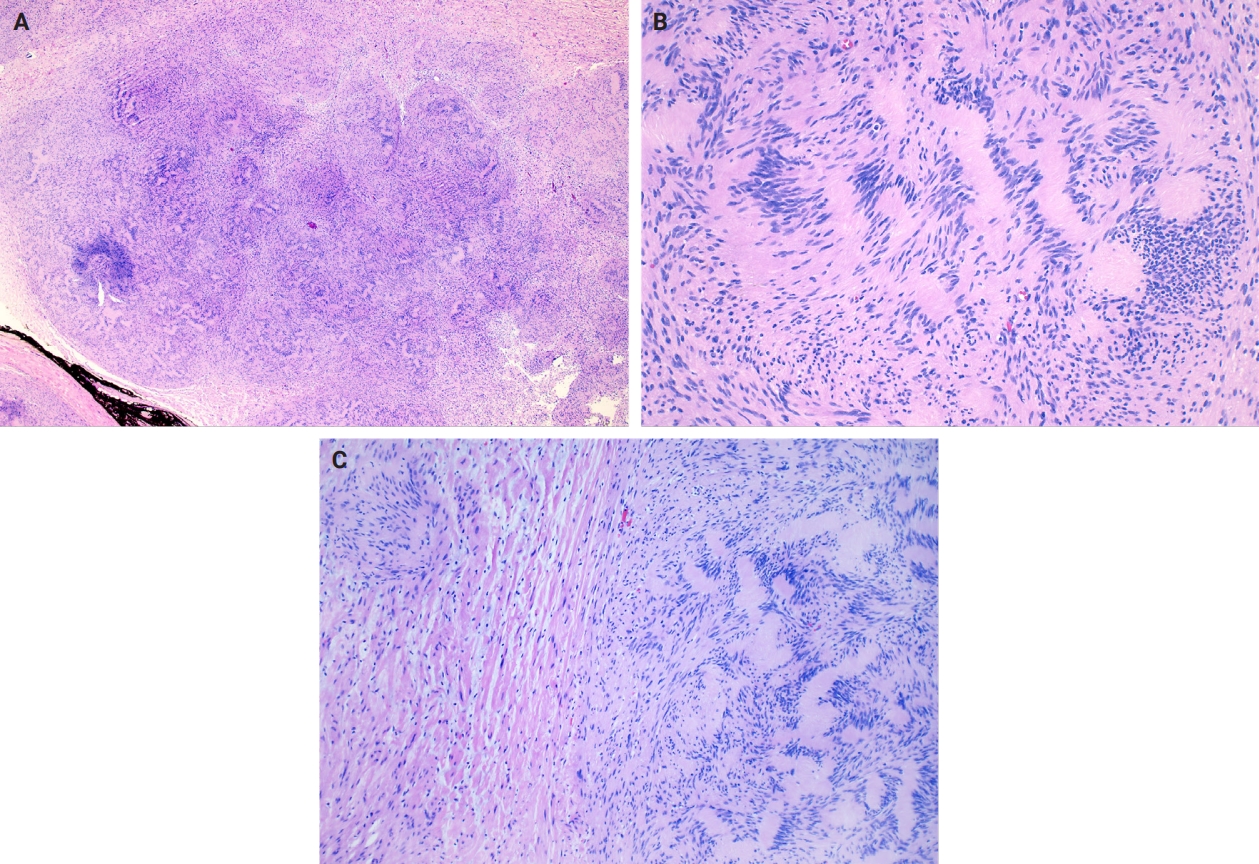
Excision of breast schwannoma with nodularity (A), Verocay bodies (B), and Antoni A and B regions (C).
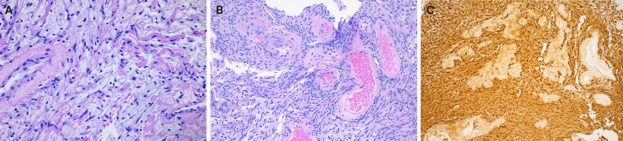
Core biopsy of breast schwannoma with myxoid loose stroma (A), variably sized hyalinized vessels with intraluminal fibrin thrombi (B) and diffuse nuclear and cytoplasmic S100 reactivity in lesional Schwann cells (S100 immunohistochemistry) (C).
It is worthwhile to be aware of the schwannoma variants, which can cause diagnostic confusion due to the histologic variations. To briefly describe, ancient schwannomas often show degenerative atypia, calcifications, cystic degeneration and various degrees of hemorrhage [20]. Cellular schwannomas have areas with a predominant Antoni A pattern, fascicular growth, lymphoid aggregates, increased mitotic activity and infiltrate margins [20]. Plexiform schwannomas have intraneural-nodular pattern, rare mitosis, and are less well-circumscribed [20]. Epithelioid cell schwannomas are characterized with epithelioid cells displaying abundant eosinophilic cytoplasm, nuclear pseudoinclusions and prominent nucleoli [19]. Reticular schwannoma is a rare variant that displays abundant myxoid change and microcysts [20]. The full profile of each variant is described in Table 1.
Immunohistochemistry
Immunohistochemistry is often needed to diagnose schwannomas, especially with small core tissue samples in the breast. The two most important positive stains are S100 (Fig. 4C) and SOX10, which are strong and diffuse in Schwann cells [3]. Schwannomas can also be positive for CD34 (weak, variable), calretinin, CD56, CD68 [21], podoplanin and Type IV collagen [22]. Classically, these are negative for estrogen receptor (ER), progesterone receptor, human epidermal growth factor receptor 2, smooth muscle markers (smooth muscle actin, desmin) and epithelial membrane antigen (capsule/perineurium only) [3]. Cytokeratins are generally negative, but may have rare labeling [23].
Cytomorphology
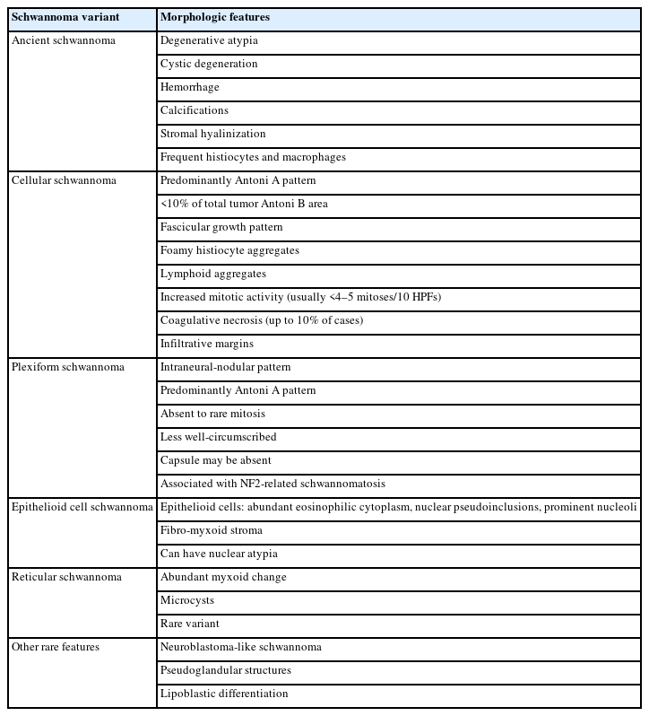
Fine needle aspiration (FNA) of breast masses is performed worldwide, but less often in the United States, where core needle biopsy (CNB) is most utilized. Cytomorphologic features of schwannoma are similar to those of other anatomic sites. These include Antoni A areas with cohesive fascicles with variable cellularity, dense fibrillary matrix, and Verocay bodies. Antoni B areas have short spindle/wavy cells, myxoid background and microcysts (Fig. 5A, B). It is important to note the limitations for utilizing FNA to diagnose schwannomas. These specimens often have low diagnostic sensitivity (0%–40%), and are often unsatisfactory due to prominent cystic change, dense stroma, and hypocellular areas. CNB has now gained acceptance as a complementary method to FNA, which provides tissue for immunohistochemical studies [24].
Fine needle aspiration of schwannoma with hypercellular, cohesive fragments of bland spindled cells (Diff-Quik) (A) and delicate fibrillary cytoplasm in fibrous stroma (Pap stain) (B).
Differential diagnosis
The differential for schwannomas is similar to other anatomic sites, including neurofibroma, neuroma, leiomyoma, nodular fasciitis, desmoid-type fibromatosis, malignant peripheral nerve sheath tumor, melanoma and dermatofibrosarcoma protuberans. However, it is important to note additional differential diagnoses particularly relevant in the breast.
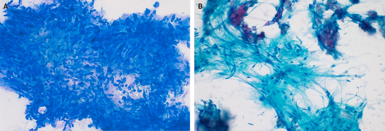
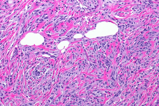
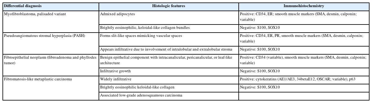
Myofibroblastoma with classic features has bland spindle cells with amphophilic cytoplasm and bright eosinophilic collagen bundles and admixed adipocytes (Fig. 6). However, the palisaded variant has spindle cells with nuclear palisading and alternating cellularity, which resembles Verocay bodies and Antoni A and B regions, respectively (Fig. 7A). These may also contain gaping, hyalinized blood vessels (Fig. 7B), cystic degeneration, and myxoid stroma. On the contrary with schwannomas, myofibroblasts have strong nuclear immunoreactivity for ER (Fig. 7C) and membranous CD34 (Fig. 7D) [25,26]. Fibroepithelial neoplasms (fibroadenoma and phyllodes tumor) may contain stromal fascicular pseudoangiomatous stromal hyperplasia (PASH)–like pattern with nuclear palisading (Fig. 8A). The lesional myofibroblasts may display elongated nuclei with palisading, resembling Verocay bodies of schwannoma (Fig. 8B) [3]. Fibromatosis-like metaplastic carcinoma is infiltrative, with fascicles of spindled cells displaying only mild to moderate cytologic atypia, including hyperchromasia and increased mitotic activity [3]. This entity is positive for a broad spectrum of cytokeratins, while schwannomas only have rare labeling [23]. PASH is hormone-dependent, which leads to an increase in stromal myofibroblasts which dissect through dense stroma. The myofibroblasts are spindled in morphology with tapering ends and occasionally wavy nuclei. Fascicular PASH has increased stromal cellularity with nuclear palisading, which overlaps in morphology with schwannoma [3]. A summary of key differentiating features of these entities is described in Table 2.
Resection of myofibroblastoma with classic features, including bland spindled cells with an amphophilic cytoplasm, dense eosinophilic collagen bundles and admixed adipocytes.
Resection of myofibroblastoma, palisading variant, with hyper- and hypocellular areas (A), hyalinized and variably dilated vessels (B), myofibroblasts with diffuse estrogen receptor (ER) nuclear positivity (ER immunohistochemistry) (C) and myofibroblasts with diffuse membranous CD34 positivity (CD34 immunohistochemistry) (D).
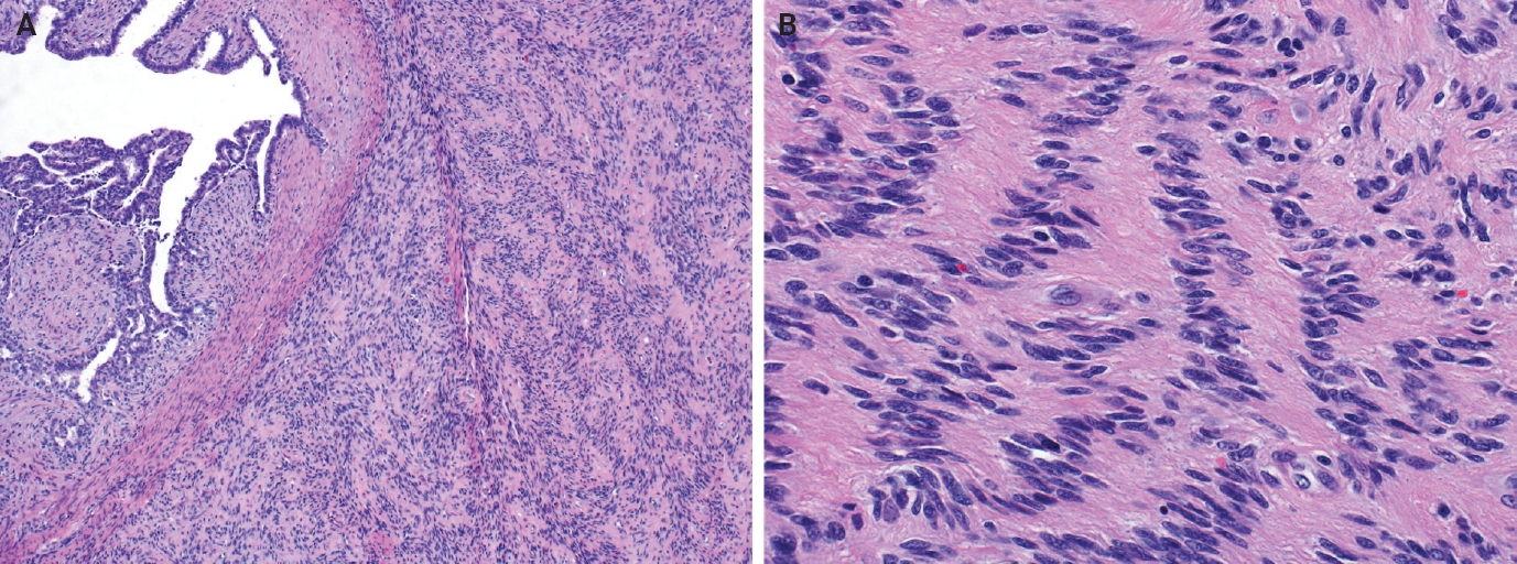
Resection of breast borderline phyllodes tumor with benign epithelial leaf-like architecture and abrupt transition to cellular stroma (A) and myofibroblasts with elongated nuclei with palisading and ribbon-like architecture, resembling Verocay bodies (B).
Mimicker neoplasms of breast schwannoma and differentiating features
CONCLUSION
Breast schwannomas have similar histologic features and immunophenotype as other anatomic sites. However, as with other breast spindle cell neoplasms, these can be particularly challenging on limited core biopsy material. Understanding histologic distinctions between the differential diagnoses and utilizing a selective immunohistochemical panel is key for classification of breast schwannomas.
Notes
Ethics Statement
Not applicable.
Availability of Data and Material
All data generated or analyzed during the study are included in this published article (and its supplementary information files).
Code Availability
Not applicable.
Author Contributions
Writing—original draft preparation: SIS, ACM. Writing—review & editing: SIS, ACM. Approval of final manuscript: all authors.
Conflicts of Interest
The authors declare that they have no potential conflicts of interest.
Funding Statement
No funding to declare.