 E-submission
Articles
- Page Path
- HOME > J Pathol Transl Med > Volume 48(3); 2014 > Article
-
Original Article
Extrapulmonary Lymphangioleiomyoma: Clinicopathological Analysis of 4 Cases - Dae Hyun Song, In Ho Choi, Sang Yun Ha, Kang Min Han, Jae Jun Lee, Min Eui Hong, Yoon-La Choi, Kee-Taek Jang, Sang Yong Song, Chin A Yi1, Joungho Han
-
Korean Journal of Pathology 2014;48(3):188-192.
DOI: https://doi.org/10.4132/KoreanJPathol.2014.48.3.188
Published online: June 26, 2014
Department of Pathology, Samsung Medical Center, Sungkyunkwan University School of Medicine, Seoul, Korea.
1Department of Radiology, Samsung Medical Center, Sungkyunkwan University School of Medicine, Seoul, Korea.
- Corresponding Author: Joungho Han, M.D. Department of Pathology, Samsung Medical Center, Sungkyunkwan University School of Medicine, 81 Irwon-ro, Gangnam-gu, Seoul 135-710, Korea. Tel: +82-2-3410-2800, Fax: +82-2-3410-0025, 'hanjho@skku.edu'
• Received: January 15, 2014 • Revised: April 13, 2014 • Accepted: April 18, 2014
© 2014 The Korean Society of Pathologists/The Korean Society for Cytopathology
This is an Open Access article distributed under the terms of the Creative Commons Attribution Non-Commercial License (http://creativecommons.org/licenses/by-nc/3.0/) which permits unrestricted non-commercial use, distribution, and reproduction in any medium, provided the original work is properly cited.
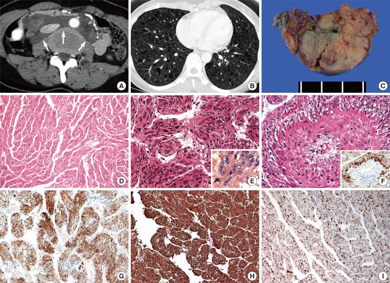
Figure & Data
References
Citations
Citations to this article as recorded by 
- Lymphangioleiomyomatosis with Tuberous Sclerosis Complex—A Case Study
Aleksandra Marciniak, Jolanta Nawrocka-Rutkowska, Agnieszka Brodowska, Andrzej Starczewski, Iwona Szydłowska
Journal of Personalized Medicine.2023; 13(11): 1598. CrossRef - A case of lymphangioleiomyomatosis with endometrial cancer diagnosed by retroperitoneoscopic para-aortic lymph node dissection
Aiko Ogasawara, Shogo Yamaguchi, Hiroaki Inui, Mieko Hanaoka, Daisuke Shintani, Sho Sato, Masanori Yasuda, Akira Yabuno
JAPANESE JOURNAL OF GYNECOLOGIC AND OBSTETRIC ENDOSCOPY.2022; 38(1): 158. CrossRef - Primary retroperitoneal PEComa: an incidental finding
Bárbara Monteiro Marinho, António Gâmboa Canha, Donzília Sousa Silva, José Davide Pinto Silva
BMJ Case Reports.2022; 15(11): e250466. CrossRef - Imaging Findings of Thoracic Lymphatic Abnormalities
Jingshuo (Derek) Sun, Thomas Shum, Fardad Behzadi, Mark M. Hammer
RadioGraphics.2022; 42(5): 1265. CrossRef - Extrapulmonary uterine lymphangioleiomyomatosis (LAM) and dysfunctional uterine bleeding: the first presentation of LAM in a tuberous sclerosis complex patient
Lucy Grant, Saliya Chipwete, San Soo Hoo, Anjali Bhatnagar
BMJ Case Reports.2019; 12(2): e226358. CrossRef - Summary of the Japanese Respiratory Society statement for the treatment of lung cancer with comorbid interstitial pneumonia
Takashi Ogura, Nagio Takigawa, Keisuke Tomii, Kazuma Kishi, Yoshikazu Inoue, Eiki Ichihara, Sakae Homma, Kazuhisa Takahashi, Hiroaki Akamatsu, Satoshi Ikeda, Naohiko Inase, Tae Iwasawa, Yuichiro Ohe, Hiromitsu Ohta, Hiroshi Onishi, Isamu Okamoto, Kazumasa
Respiratory Investigation.2019; 57(6): 512. CrossRef - Incidental lymphangioleiomyomatosis in the lymph nodes of gynecologic surgical specimens
Ikumi Kuno, Hiroshi Yoshida, Hanako Shimizu, Takashi Uehara, Masaya Uno, Mitsuya Ishikawa, Tomoyasu Kato
European Journal of Obstetrics & Gynecology and Reproductive Biology.2018; 231: 93. CrossRef - Solitary extrapulmonary lymphangioleiomyomatosis of the liver: A case report and literature review
Weiwei Fu, Yujun Li, Hong Li, Ping Yang, Xiaoming Xing
Experimental and Therapeutic Medicine.2016; 12(3): 1499. CrossRef - Incidental Pelvic and Para-aortic Lymph Node Lymphangioleiomyomatosis Detected During Surgical Staging of Pelvic Cancer in Women Without Symptomatic Pulmonary Lymphangioleiomyomatosis or Tuberous Sclerosis Complex
Joseph T. Rabban, Brandie Firetag, Ankur R. Sangoi, Miriam D. Post, Charles J. Zaloudek
American Journal of Surgical Pathology.2015; 39(8): 1015. CrossRef
 PubReader
 Cite this Article

