Ovarian Remnant Syndrome at the Trochar Site: A Report of a Rare Complication Following Laparoscopic Ovarian Surgery
Article information
Ovarian surgery can leave behind ovarian tissue and this residual ovarian tissue may eventually cause pelvic pain and/or the formation of a mass. The term "ovarian remnant syndrome" has been used to describe this condition.1 Ovarian remnant syndrome usually occurs as a result of improper tissue removal or an inappropriate blunt dissection. Endometriosis, pelvic inflammatory disease or previous gynecologic surgeries have been known to increase the risk. Such conditions make removal of ovarian tissue difficult, due to the increased likelihood of dense fibrotic adhesions between an ovary and surrounding structures. Furthermore, with an increase in the number of laparoscopic ovarian surgeries performed, implantation of ovarian tissue also has been recognized as an important cause of ovarian remnant syndrome.2
Various locations have been identified where residual ovarian tissue was detected after laparoscopic ovarian surgery, including the pelvic wall, cervix, vagina, and bladder.1 However, reports of residual ovarian tissue detected in an abdominal wall have been rare. This report describes a case of ovarian remnant syndrome caused by residual ovarian tissue at the trochar site after a laparoscopic ovarian cystectomy.
CASE REPORT
A 35-year-old woman presented with an abdominal wall mass that was palpable one week prior to her visit. The mass was not painful and did not show a change in size. Her past medical history was remarkable because she had undergone laparoscopic bilateral ovarian cystectomy due to endometriosis 3 years prior to her visit. Physical examination revealed a surgical scar on her left lower abdomen and an index finger tip-sized mass was palpable in the abdominal wall under the scar. It was smooth and movable, and was not tender. The laboratory findings were unremarkable.
Ultrasonography demonstrated an approximately 1.6×0.9 cm-sized hypoechoic mass in the subcutaneous adipose tissue (Fig. 1). An excisional biopsy was performed. The mass was well demarcated from the adjacent soft tissue by a thin fibrous capsule. The cut surface showed a yellow, glistening lobular parenchyma with areas of blood-filled cystic spaces. Histologically, the parenchyma was composed of luteinized granulosa and theca cell layers, suggesting corpus luteum of the ovary (Fig. 2). There was no evidence of endometriosis.
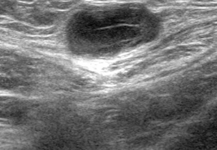
Ultrasonography demonstrats a hypoechoic mass in the abdominal wall under the trochar site.
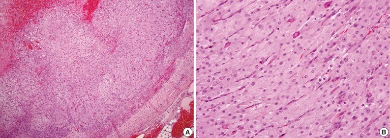
Histologically, the mass was surronded by thin fibrous capsule and showed intraparenchymal hemorrhaae (A). The parenchyma consisted of hyalinized granulosa cells and theca cells (B).
A review of the operation record of the laparoscopic surgery that was performed 3 years prior showed that the patient had 5.4×4.5×4.0 cm- and 3.2×3.0×1.5 cm-sized endometriotic cysts in bilateral ovaries with an associated dense pelvic adhesion. After a cystectomy, the specimens were fragmented and extracted using forceps through the trochar on her left lower abdomen.
The patient was discharged after the abdominal wall mass excision and was healthy with no recurrence of the lesion at a follow-up at 32 months.
DISCUSSION
Ovarian remnant syndrome is a rare but gradually increasing complication after ovarian surgery. Patients with ovarian remnant syndrome often experience chronic constant or cyclic pelvic pain. As a diagnostic method, a clomiphen citrate stimulation test can be helpful. This test elicits ovarian follicular development in residual ovarian tissues and the structure change can be observed by serial ultrasonographic examination. Residual ovarian tissue has shown various histological spectra, such as normal ovarian cortex, corpus luteum, endometriosis and rarely, malignant transformation. The therapeutic choice of ovarian remnant syndrome is surgical resection of residual ovarian tissue. It is mostly curative and also prevents the possibility of malignant transformation. Therefore, ovarian remnant syndrome is an important differential diagnosis that should be considered and managed in patients with pelvic pain who underwent oophorectomy.1
Several theories have been suggested to explain the occurrence of ovarian remnant syndrome during laparoscopic surgery, of which, implantation of ovarian tissue is the most important.2 Several experimental studies performed artificial implantation of ovarian tissue in animals. These studies have shown that implanted ovarian tissue can be revascularized without artificial vascular anastomosis and still be viable. Several growth factors such as fibroblast growth factor, transforming growth factor and vascular endothelial growth factor have been found to play a role in this process. Eventually, implanted ovarian tissue can undergo a normal ovarian cycle with a response to estrogen and progesterone.3
Most cases of ovarian remnant syndrome after laparoscopic ovarian surgery have been caused by residual ovarian tissue in a pelvic cavity. To our knowledge, there have been only two cases that showed residual ovarian tissue on an abdominal wall. In the previous and present cases, clinical presentation, indication of previous surgery and histological findings of residual ovarian tissue were relatively typical (Table 1).
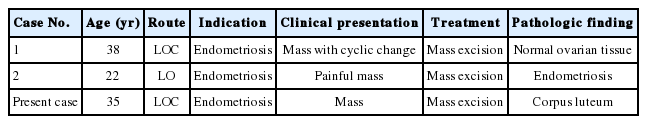
Cases of ovarian autograft in abdominal wall after laparoscopic ovarian surgery
In the case described by Marconi et al.,4 a piece of ovarian tissue remained in the patient's abdominal wall during operation. The author suggested that this piece was the source of residual ovarian tissue. In the case by Chao5 as well as the present case, the source of residual ovarian tissue was not determined but implantation of a tiny piece of ovarian tissue might occur during the extraction of the specimen through the trochar.4 These cases suggest that during specimen extraction after laparoscopic ovarian surgery, the risk of ovarian tissue implantation on the abdominal wall should be recognized and prevented. Several preventive methods have been suggested and include the use of large-diameter trochars, copious irrigation of the operation field, plastic bags and a vaginal approach with culdotomy.4,5
In conclusion, we report a rare complication of laparoscopic ovarian surgery: implantation of ovarian tissue on the abdominal wall. The present case suggests that the risk of ovarian tissue implantation during extraction of the specimen after laparoscopic ovarian surgery should be recognized. In patients with ovarian remnant syndrome, surgical resection of residual ovarian tissue is mandatory and therefore, it is wiser to prevent the occurrence of such conditions.
Notes
No potential conflict of interest relevant to this article was reported.