Myoferlin Expression and Its Correlation with FIGO Histologic Grading in Early-Stage Endometrioid Carcinoma
Article information
Abstract
Background
For endometrioid carcinoma patients, International Federation of Gynecologists and Obstetricians (FIGO) histologic grading is very important for identifying the appropriate treatment method. However, the interobserver discrepancy with this three-tiered grading system is a serious potential problem. In this study, we used immunohistochemistry to analyze the relationship between FIGO histologic grading score and myoferlin expression.
Methods
We studied the endometrioid carcinoma tissues of 60 patients from Gyeongsang National University Hospital between January 2002 and December 2009. Immunohistochemical analysis of myoferlin was performed on tissue microarray blocks from surgical specimens.
Results
Myoferlin expression was observed in 58 of 60 patients. Moderate and strong myoferlin expression was observed in low-grade endometrioid carcinoma, while there was a tendency toward loss of myoferlin expression in high-grade endometrioid carcinoma (p<.001).
Conclusions
Our study revealed that myoferlin loss is significantly correlated with high FIGO grade of endometrioid carcinoma.
In endometrioid carcinoma patients, International Federation of Gynecologists and Obstetricians (FIGO) histologic grading is very important in determining the treatment method and predicting the patient’s prognosis [1-3]. Particularly, it is recommended that patients with FIGO stage I and histologic grade 3 endometrioid carcinoma undergo adjuvant radiotherapy after surgery [3], while patients with FIGO stage I and histologic grade 1 or 2 endometrioid carcinoma should be treated with surgery only. However, the interobserver discrepancy of this three-tiered FIGO grading system can pose a serious potential problem. Thus, many authors have used immunohistochemical (IHC) staining [4-6], genetic molecular testing [7-9], and other two-tiered systems [10,11] to reduce the discrepancy.
Myoferlin, a protein in the cellular membrane, is involved in cellular regeneration after injury [12]. Recent studies have reported a correlation between the prognosis of cancer patients and myoferlin expression in breast, lung, oropharyngeal, and pancreatic cancer [13-16].
In this study, we used IHC staining to analyze the relationship between FIGO histologic grading and myoferlin expression.
MATERIALS AND METHODS
Case selection
We collected clinical data from the charts of endometrioid carcinoma patients treated at Gyeongsang National University Hospital, Jinju, Korea, between January 2002 and December 2009. A total of 60 patients who underwent hysterectomy were enrolled. The tumor stage and histologic grade of each case were assessed using the FIGO system. All gross photographs and hematoxylin and eosin–stained glass slides of surgical specimens were reviewed by two pathologists.
This study was approved by the Institutional Review Board of Gyeongsang National University Hospital with a waiver of informed consent (IRB No. GNUH-2015-12-003).
Tissue microarray
Tumor samples were fixed overnight in 20% neutral-buffered formalin and were examined grossly, dissected, and embedded in paraffin blocks. One or two representative blocks were selected by microscopic examination. One representative core (3 mm in diameter) tissue was obtained from each paraffin block and arranged in new recipient tissue microarray blocks. Representative areas of the donor blocks were selected from near the invasive front.
IHC analysis
A primary antibody for myoferlin (1:100, 7D6, Abcam, Cambridge, UK) was used to investigate protein expression. The IHC method used was described in detail in our previous report [14].
The positive control for myoferlin was normal endometrial tissue. Tissues showing an intensity equal to or stronger than that of normal endometrial tissue were classified as grade 3. The IHC slides were scored using the three-tiered FIGO system by two pathologists.
Statistical analysis
Correlation analyses were performed using the chi-square test. A p-value of < .05 was considered statistically significant. All statistics were analyzed using SPSS ver. 24.0 (IBM Corp., Armonk, NY, USA).
RESULTS
Clinicopathological features of the patients
The clinicopathological features of 60 endometrioid carcinoma patients are summarized in Table 1. The mean age was 51 years. All patients underwent hysterectomy. Among 60 cases, 43 (71.7%) were classified as FIGO stage Ia after surgery, whereas 40 (66.7%) were considered FIGO histologic grade 1.
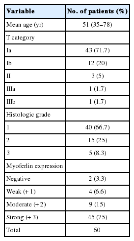
Clinical and pathological features of 60 endometrioid carcinoma patients
Myoferlin expression in nontumorous endometrial tissue
Nontumorous endometrial tissues were obtained from leiomyoma or adenomyosis patients who underwent hysterectomy. In normal epithelial tissues, moderate and strong myoferlin expression was observed. Specifically, endometrial tissue in secretory phase showed strong expression (Fig. 1).
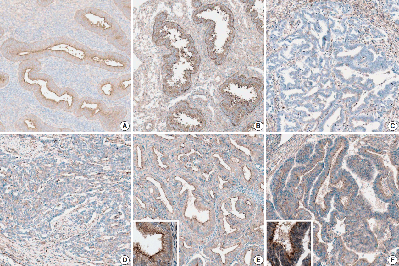
Myoferlin expression in nontumorous endometrial tissue in proliferative phase (A) and secretory phase (B). (C) Loss of myoferlin expression in high-grade endometrioid carcinoma. Weak (+1, International Federation of Gynecologists and Obstetricians [FIGO] grade 3) (D), moderate (+2, FIGO grade 2) (E), and strong (+3, FIGO grade 1) (F) expression of myoferlin in endometrioid carcinoma.
Myoferlin expression in endometrioid carcinoma
The clinicopathological features of the cases included in this study are summarized in Table 1. Myoferlin expression in the cytoplasm and cellular membrane of cancer cells was observed in 58 of the 60 patients (Fig. 1). Similarly, cytoplasmic and membranous expression of myoferlin was evident in normal endometrial glands in proliferative and secretory phases. Forty-five patients (75%) exhibited strong myoferlin expression, similar to that in normal endometrial glands.
Correlation between myoferlin expression and FIGO histologic grading
The correlation is described in Table 2. In the chi-square test, moderate and strong myoferlin expression was observed in low-grade endometrioid carcinoma, while there was a tendency toward loss of myoferlin expression in high-grade endometrioid carcinoma (p < .001).
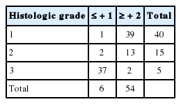
Correlation between myoferlin expression and FIGO histologic grading
Correlation between myoferlin expression and FIGO staging
The correlation is shown in Table 3. In the chi-square test, most patients with FIGO stage III endometrioid carcinoma exhibited negative or weak myoferlin expression (+ 1). A tendency toward loss of myoferlin expression was also observed in late-stage endometrioid carcinoma (p < .001).
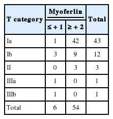
Correlation between myoferlin expression and FIGO staging
DISCUSSION
Myoferlin is a little known protein. To our knowledge, this study is the first to identify myoferlin expression in endometrial tissue. Recent studies have reported that myoferlin contributes to the proliferation, migration, and invasion of cancer cells and is overexpressed in several types of cancer. However, in this study, endometrial cancer showed the opposite result; myoferlin expression was decreased in high-grade endometrioid carcinoma, probably because normal endometrial tissue undergoes a continuous cycle of regeneration. Moreover, myoferlin has been reported to be involved in cellular regeneration after injury [14]. The tumorigenesis of endometrioid carcinoma is associated with noncyclic continuous exposure to sex hormones. Our results implicate a correlation among cellular regeneration, hormonal effect, and myoferlin expression.
Though FIGO grading is important in guiding patient treatment, the interobserver discrepancy of this three-tiered grading system is a potential problem. Hence, many authors have attempted grading using IHC staining [4-6], genetic molecular testing [7-9], other two-tiered systems [10,11], and curettage and cytology [17] to decrease discrepancies.
In a previous study, Daniilidou et al. [5] investigated PTEN and p53 gene expression in endometrioid and serous papillary carcinoma and showed that these biomarkers contribute to accurate diagnosis and therapeutic decisions in relation to tumor stage and grade. In another notable study on the cytologic scoring of endometrioid carcinoma of the endometrium, Nishimura et al. [17] examined 64 cytologic samples and scored them using 10 cytology characteristics. The cytologic grade was closely related to histologic grade, and a high cytologic score was correlated with p53 mutation and myometrial invasion. The study implied that the cytologic scoring system for endometrioid carcinoma is useful for predicting histologic grade and malignant potential of the tumor.
Guan et al. [10] tried a new binary grading system for endometrial carcinoma and compared it with an existing binary grading system and FIGO grading in hysterectomy specimens. They examined 254 hysterectomies and graded them according to the new grading system including architecture pattern and nuclear atypia. They concluded that the three-tiered FIGO grading system retained superior prognostic power. However, the new binary grading system is an attractive option due to its good reproducibility and the elimination of ambiguity of intermediate grades.
In the present study, we also tried to decrease discrepancies by determining the FIGO grade according to myoferlin expression. Our statistically significant (p < .001) finding was that moderate and strong myoferlin expression was observed in low-grade endometrioid carcinoma, and loss of myoferlin expression was noted in high-grade endometrioid carcinoma. Here, we found that myoferlin could be a valuable marker for the accurate grading of uterine endometrioid carcinoma.
In this study, the level of myoferlin expression in endometrioid carcinoma was opposite that observed in other studies, suggesting a hidden mechanism underlying the continuous regeneration of tissue in the endometrium. Thus, further investigation in the role of myoferlin in the endometrium and in tumorigenesis of endometrioid carcinoma is recommended.
This study had some limitations. The number of patients involved was only 60, and the cases consisted of mostly early-stage endometrioid carcinoma. Therefore, further studies with larger sample sizes are necessary to validate the significance of myoferlin expression in early- and late-stage endometrioid carcinoma.
In conclusion, our study revealed that myoferlin loss is significantly correlated with high FIGO grade of endometrioid carcinoma. To our knowledge, this is the first report on myoferlin expression in endometrial tissue, and our results could help in the management of patients with endometrioid carcinoma.
Notes
Conflicts of Interest
No potential conflict of interest relevant to this article was reported.
Acknowledgements
This work was supported by the Development Fund Foundation, Gyeongsang National University (2015).