Extra-axial poorly differentiated chordoma of the knee masquerading as periprosthetic joint infection: a case report
Article information

Abstract
Poorly differentiated chordoma (PDC) is a malignant neoplasm defined by the co-expression of brachyury and cytokeratin with loss of SMARCB1 (INI-1) expression. Extra-axial PDC is exceedingly rare, with only 10 cases reported to date. Here, we report an additional case of extra-axial PDC arising in the soft tissues surrounding the knee joint of a 69-year-old woman, which was initially misdiagnosed as periprosthetic joint infection following total knee arthroplasty. Histologically, the tumor comprised epithelioid to spindled cells with vacuolated, eosinophilic cytoplasm, focal rhabdoid areas, without myxoid stroma. Immunohistochemically, the tumor cells showed diffuse positivity for brachyury and cytokeratin, with loss of INI-1 expression. Next-generation sequencing identified a homozygous deletion of CDKN2A, but no SMARCB1 genomic alteration was detected despite INI-1 loss. This case shows that extra-axial PDC should be considered in the differential diagnosis of soft tissue tumors, irrespective of anatomical site or morphology.
INTRODUCTION
Chordoma is a rare malignant neoplasm exhibiting notochordal differentiation [1]. A defining molecular feature is the expression of brachyury, a transcription factor essential for notochordal development encoded by the TBXT gene on chromosome 6q27 [2]. Chordoma arises predominantly along the axial skeleton, with predilection for the skull base, vertebral bodies, and sacrococcygeal region [3]. According to the current World Health Organization classification of tumors, chordoma is divided into three subtypes: conventional, dedifferentiated, and poorly differentiated chordoma (PDC). Among these, PDC is a rare entity that predominantly affects children and young adults and carries a poorer prognosis than conventional chordoma. It is defined by loss of SMARCB1 (INI-1) expression in the setting of retained notochordal markers, including brachyury and cytokeratin [4]. Histologically, PDC consists of sheets of epithelioid cells, frequently with focal rhabdoid morphology. Unlike conventional chordoma, PDC characteristically lacks the myxoid stroma and physaliphorous cells [3].
Occurrence outside the axial skeleton is exceedingly rare, with only 10 cases of extra-axial PDC reported to date [5-9]. Its atypical anatomical location and nonspecific histomorphological features, which overlap with a broad spectrum of malignancies, mean that extra-axial PDC is not commonly included among the initial diagnostic considerations. Here, we report a case of extra-axial PDC arising at the knee joint, illustrating the diagnostic challenges posed by this uncommon entity in an atypical anatomical setting.
CASE REPORT
The patient is a 69-year-old female who had previously undergone staged bilateral total knee arthroplasty (TKA) for osteoarthritis at an outside hospital; the left TKA was performed 3 months prior, followed by the right TKA 2 months prior. Postoperatively, she experienced persistent pain, swelling, and progressive joint contracture in the left knee, while the right knee remained asymptomatic. Magnetic resonance imaging (MRI) performed at the outside hospital to evaluate persistent symptoms demonstrated a mass lesion extending from the left knee joint space to the distal femur (Fig. 1). She was referred to our institution for further evaluation and management. Upon presentation, physical examination revealed localized warmth and erythema over the left knee. Based on these clinical findings and her surgical history, periprosthetic joint infection was the primary clinical suspicion. During surgery for presumed infection, whitish tissue resembling granulation tissue or rice bodies was noted. This appearance raised the intraoperative suspicion of tuberculous infection, and tissue specimens were submitted for histopathological examination and microbial cultures. The prosthesis was explanted, followed by irrigation and placement of an antibiotic-loaded cement spacer.
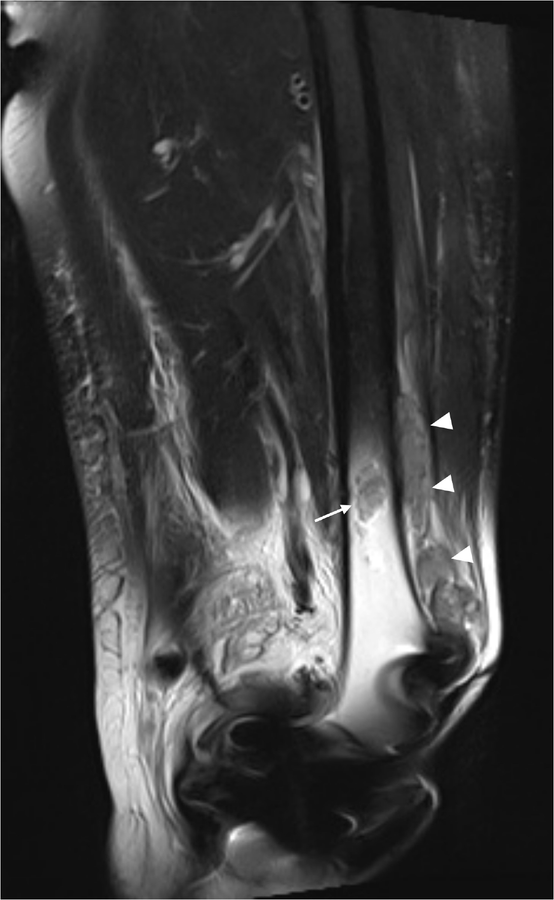
Magnetic resonance imaging of the left knee. The mass extends from the superior joint space through the pre-femoral fat pad to the anterior parosteal region of the distal femoral shaft (arrowheads). A separate 3.2 cm lesion with central marrow involvement is identified in the left distal femoral shaft (arrow).
Radiologic findings
Computed tomography (CT) revealed a lobulated, ill-defined hypodense mass measuring 14 cm in greatest dimension, with peripheral rim enhancement. The mass extended from the superior joint space of the left knee through the pre-femoral fat pad to the anterior parosteal region of the distal femoral shaft. MRI demonstrated a heterogeneous soft tissue mass with T2 low-to-intermediate signal intensity and periosteal involvement, accompanied by a large joint effusion and focal hemorrhage. A separate 3.2 cm lesion with central marrow involvement was identified in the left distal femoral shaft, suspicious for regional bone metastasis (Fig. 1). 18F-fluorodeoxyglucose positron emission tomography (18F-FDG PET)/CT revealed intense hypermetabolic activity in the left knee mass and distal femur, with additional hypermetabolic lesions in the left inguinal, external iliac, and common iliac lymph nodes, as well as the right pelvic bone, right distal femur, and hepatic segments 5 and 6, suggestive of metastatic disease.
Microscopic findings
Macroscopically, the biopsy specimen was whitish-tan and showed a multinodular appearance (Fig. 2). Microscopically, the lesion showed an infiltrative, multilobular growth pattern, extending into the adjacent muscle. The tumor cells were arranged in sheets and nests, composed of epithelioid to spindled cells with abundant vacuolated and eosinophilic cytoplasm (Fig. 3A–C). The nuclei showed coarse chromatin with prominent nucleoli and frequent mitotic activity (Fig. 3D). Multiple foci of necrosis and focal multinucleated tumor giant cells were identified within the tumor sheets and nests. Focal rhabdoid cells were also identified, accounting for less than 5% of the total tumor volume (Fig. 3E). Dense lymphocytic infiltrates were present at the periphery of the tumor nests, accompanied by hemosiderin deposition (Fig. 3F). Myxoid stroma was entirely absent, and no conventional chordoma component, defined by cords or chains of cells within a myxoid matrix, was identified. Based on the microscopic findings, the initial differential diagnostic considerations included clear cell sarcoma, metastatic clear cell renal cell carcinoma, epithelioid sarcoma, synovial sarcoma, and malignant melanoma. To further characterize the lesion, a comprehensive immunohistochemical (IHC) study was performed. The tumor cells showed diffuse positivity for pan-cytokeratin (pan-CK) (Fig. 4A) and epithelial membrane antigen (EMA). S100 protein was focally positive (Fig. 4B), as were cytokeratin (CK) 7 and CK20. Complete loss of INI-1 nuclear expression was observed (Fig. 4C), together with diffuse nuclear positivity for brachyury (Fig. 4D). These findings were consistently observed across all morphologic components, including the focal rhabdoid areas. The Ki-67 labeling index was estimated at 55.1%. Immunostaining for p53 showed a wild-type expression pattern. The tumor was negative for all other markers tested, including CD68, CD30, anaplastic lymphoma kinase, CD3, CD20, SOX10, HMB45, paired box 8 (PAX8), CD10, carbonic anhydrase IX (CAIX), transducin-like enhancer of split 1 (TLE1), SS18-SSX, thyroid transcription factor 1, ERG, CD34, calponin, spalt-like transcription factor 4 (SALL4), and special AT-rich sequence-binding protein 2 (SATB2). This immunoprofile, characterized by co-expression of brachyury and cytokeratin with loss of INI-1, confirmed the diagnosis of poorly differentiated chordoma arising at an extra-axial location.
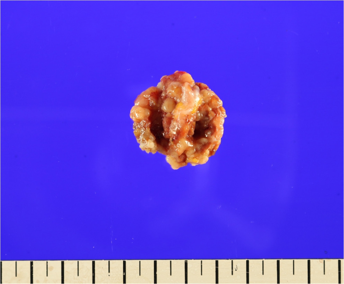
Gross photograph of the biopsy specimen demonstrates a whitish-tan and multinodular appearance.
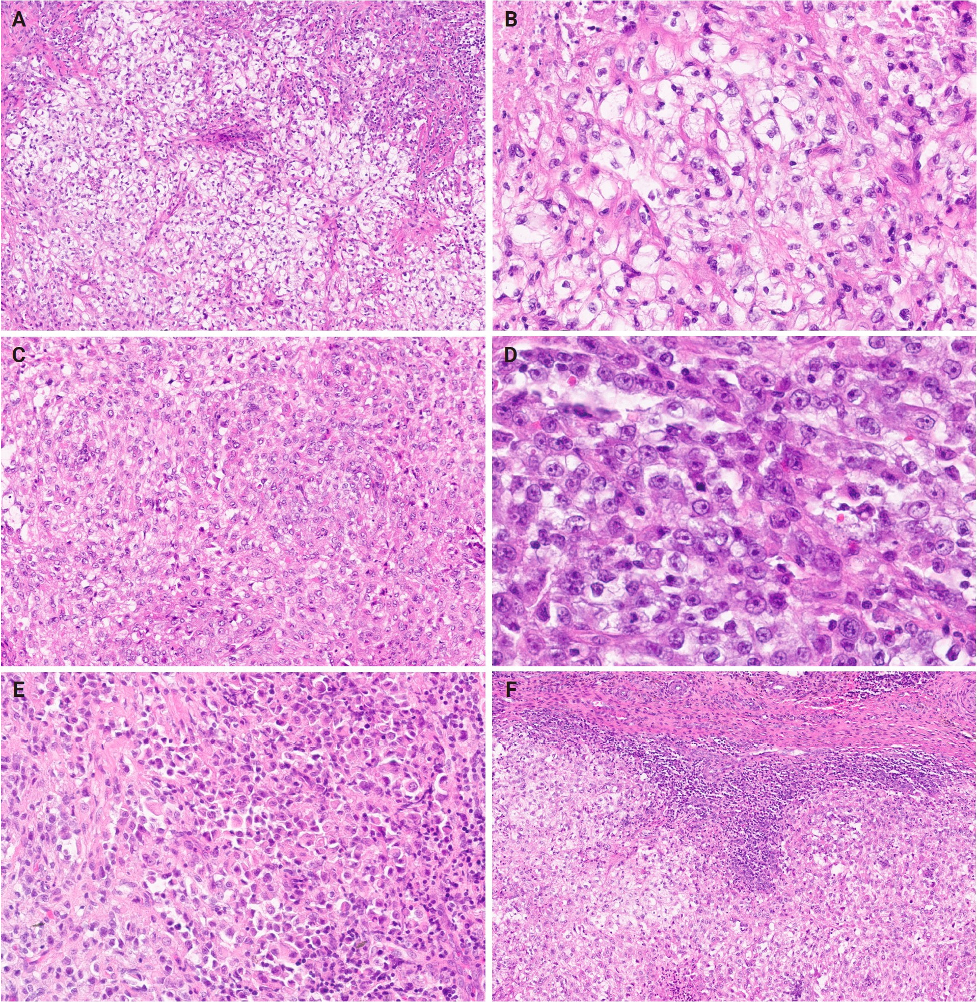
Microscopic findings of the tumor. (A) Tumor cells arranged in sheets and nests, composed of epithelioid to spindled cells. (B) A subset of tumor cells showing vacuolated cytoplasm with distinctive cell borders. (C) Tumor cells with abundant eosinophilic cytoplasm. (D) Tumor cells demonstrating coarse chromatin with prominent nucleoli and frequent mitotic activity. (E) Focal rhabdoid cells accounting for less than 5% of the total tumor volume. (F) Dense lymphocytic infiltrates at the periphery of the tumor nests.
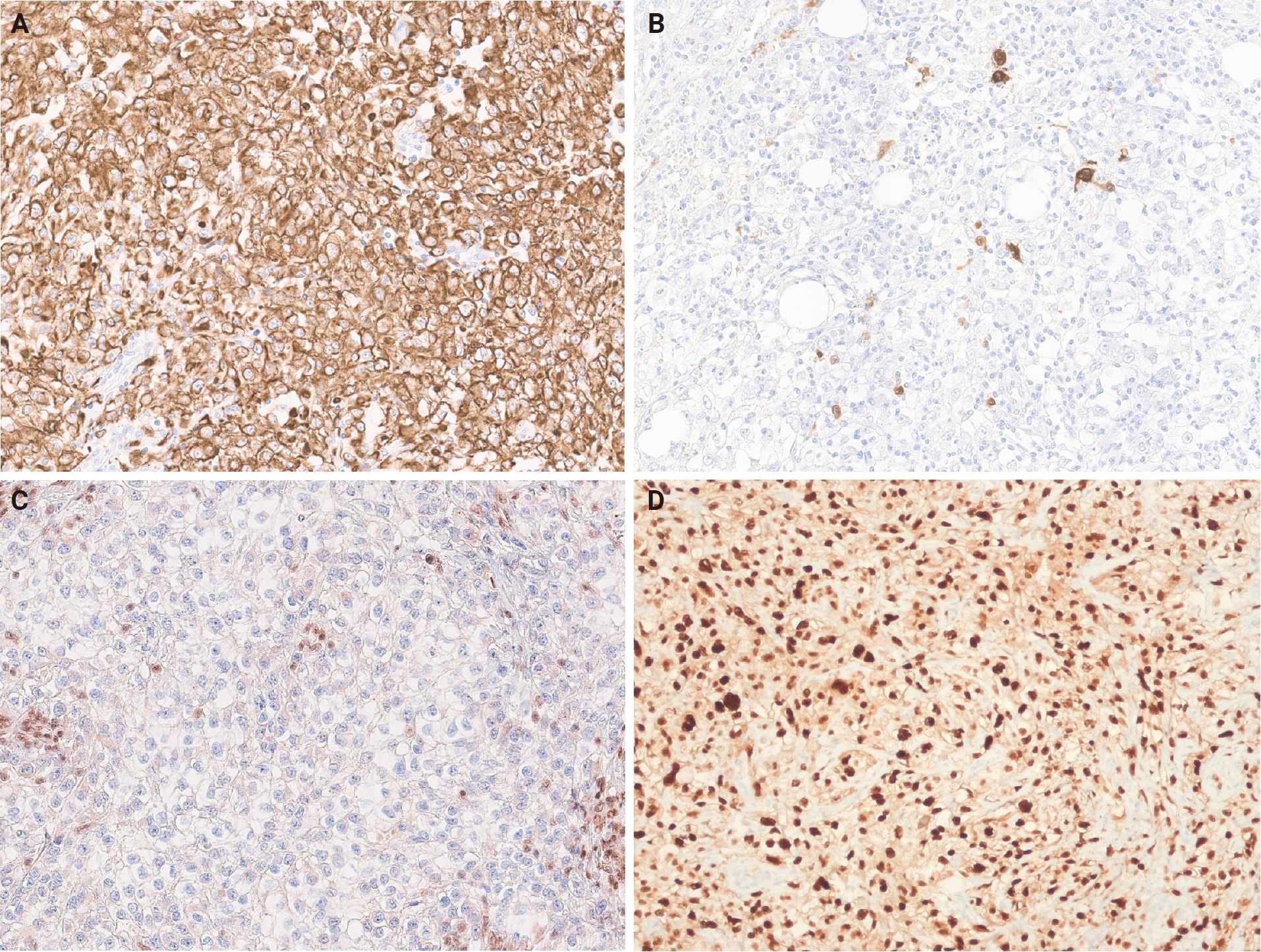
Immunohistochemical findings. (A) Diffuse positivity for pan-cytokeratin in tumor cells. (B) Focal positivity for S100 protein in tumor cells. (C) Loss of INI-1 nuclear expression in tumor cells. (D) Diffuse nuclear positivity for brachyury in tumor cells.
Molecular findings
Next-generation sequencing (NGS) identified a homozygous deletion of CDKN2A (copy number: 0.48), a nonsense mutation in ATM (c.8710G>T, p.E2904*; variant allele frequency [VAF] 56.66%), and a frameshift mutation in MSH6 (c.4068_4071dup, p.K1358Dfs*2; VAF 50.91%). Despite the loss of INI-1 protein expression on immunohistochemistry, no pathogenic genomic alterations involving the SMARCB1 gene were detected. Detailed genomic findings and additional variants of uncertain significance are summarized in the Supplementary Table S1.
Clinical course
Following the pathological diagnosis, the patient was lost to follow-up at our institution, and no further information regarding treatment or clinical outcome was available. Biopsy of the radiologically suspected metastatic lesions, including the distal femoral marrow lesion and the inguinal, external iliac, and common iliac lymph nodes, was not performed.
DISCUSSION
The present case shared the histomorphologic and IHC features characteristic of previously reported extra-axial PDC cases [5-9]. Histologically, the tumor demonstrated a multinodular, infiltrative growth pattern composed of sheets and nests of epithelioid to spindled cells with abundant vacuolated and eosinophilic cytoplasm, focal rhabdoid morphology, prominent necrosis, and a dense peripheral lymphocytic infiltrate, all of which have been described in prior reports of extra-axial PDC [8,9]. Myxoid stroma, a hallmark of conventional chordoma, was entirely absent. By immunohistochemistry, the tumor cells showed diffuse positivity for pan-CK and focal positivity for S100 protein, with complete loss of INI-1 nuclear expression and diffuse nuclear positivity for brachyury. This immunoprofile, comprising co-expression of cytokeratin and brachyury with loss of INI-1, is consistent with those of previously reported extra-axial PDC cases and formed the diagnostic cornerstone in the present case [5-9].
The differential diagnosis of this tumor was broad because of its extra-axial location, epithelioid and focal rhabdoid morphology, cytokeratin positivity, and INI-1 loss. Among INI-1–deficient neoplasms, proximal-type epithelioid sarcoma and malignant rhabdoid tumor were considered. Epithelioid sarcoma shares cytokeratin, EMA positivity, and INI-1 loss with PDC, but CD34, expressed in more than 50% of epithelioid sarcomas, was absent, and both entities lack brachyury expression [10]. Malignant rhabdoid tumor is predominantly composed of sheets of rhabdoid cells and was further excluded by negativity for brachyury and SALL4 [11]. Myoepithelial carcinoma and epithelioid malignant peripheral nerve sheath tumor were also considered given their overlapping epithelioid morphology and occasional INI-1 loss, but both characteristically express SOX10, which was absent in this case [12]. Myoepithelial carcinoma was further excluded by the absence of calponin expression, another marker of myoepithelial differentiation [13]. Clear cell sarcoma, which shows melanocytic differentiation with HMB45 and SOX10 positivity, and synovial sarcoma, which is characterized by strong TLE1 expression and SS18-SSX fusion, were both excluded as none of these features were present in this case [14,15].
Metastatic poorly differentiated carcinoma was also included in the differential diagnosis. Metastatic clear cell renal cell carcinoma, which shows a nested growth of clear cells with an arborizing vascular network, was excluded by negativity for PAX8, CD10, and CAIX [16]. To evaluate for metastatic chordoma from a primary axial site, whole-body 18F-FDG PET/CT and contrast-enhanced CT of the chest and abdomen were performed, revealing no evidence of a primary lesion in the axial skeleton, and the diffuse nuclear brachyury positivity in the knee mass further supports a notochordal origin rather than metastasis from a non-chordomatous primary; however, biopsy of the suspected metastatic lesions was not performed in this case. A recent large-scale tissue microarray study by Caneve et al. [17] analyzing 14,976 tumor samples demonstrated that brachyury can very rarely show focal weak expression in germ cell tumors and colorectal adenocarcinoma. To further exclude germ cell tumor origin, SALL4 was performed and was negative, and SATB2, expressed in colorectal adenocarcinoma, was also negative. Finally, prosthetic implant-associated sarcoma was considered in view of the patient's history of bilateral TKA. However, reported implant-associated sarcomas have a mean latency of approximately 9 years from implant placement to diagnosis [18], and the interval of only 3 months in the present case argued against this diagnosis. These findings excluded the relevant morphologic and clinical mimickers and, combined with the overall immunoprofile of the tumor, confirmed the diagnosis of extra-axial PDC.
With the diagnosis of extra-axial PDC established, we next reviewed the clinicopathologic features of previously reported cases to contextualize the present findings. To date, 10 cases of extra-axial PDC have been reported in the literature. In our case, the tumor originated in the soft tissue surrounding the knee joint with suspected secondary involvement of the femur. Including the present case, extra-axial PDC has now been described in 11 cases, among which the knee joint was the most common site, accounting for eight cases (73%). In all of these eight cases, tumors arose in the soft tissues surrounding the knee joint. One case originated as a primary intraosseous tumor in the distal femur adjacent to the knee, and the remaining two arose in the soft tissues of the wrist and elbow joints, respectively [5-9]. Among 10 soft tissue–based cases including the present case, seven cases demonstrated involvement of the adjacent bone, while the others remained confined to the soft tissue without osseous extension. Based on the accumulated cases to date, extra-axial PDC appears to have a predilection for the intra-articular space and surrounding soft tissues of joints. Why the majority of extra-axial PDC cases arise around the knee joint remains unclear, despite the fact that the extremities and their surrounding soft tissues develop independently of the notochord. However, from a developmental perspective, the extremities and joints arise from the lateral mesoderm, which itself originates from primitive streak cells that transiently express brachyury during early embryogenesis. Although brachyury expression is subsequently suppressed in fully differentiated limb and joint tissue, the transcriptional and signaling states of this early developmental phase may leave a lasting imprint on chromatin architecture. This epigenetic memory could persist even after brachyury expression is silenced in mature tissue [19]. The periarticular region is enriched with mesenchymal progenitor cells residing in structures such as the synovium and fat pad [20]. We speculate that if this dormant brachyury expression were aberrantly reactivated by environmental or genetic factors within this progenitor-rich niche, it could represent a developmental mechanism underlying the emergence of extra-axial chordoma [19,21]. In the present case, the patient's persistent pain and localized warmth following TKA led to an initial clinical suspicion of periprosthetic joint infection, which delayed the diagnosis of malignancy. Among the 10 previously reported extra-axial PDC cases, one patient was diagnosed with extra-axial PDC arising around a knee joint that had also undergone TKA [7,8]. In another case, the patient had a history of restricted knee joint movement following an intra-articular fracture, persisting for more than 50 years [6]. Our own patient also had pre-existing osteoarthritis of the knee prior to TKA. Considered together, these observations raise the possibility that chronic joint injury and the associated regenerative process may act as an environmental trigger for aberrant brachyury reactivation and SMARCB1 loss. However, this remains speculative, and the precise mechanism underlying the predilection of extra-axial PDC for the knee joint requires further investigation. These findings suggest that extra-axial PDC should be included in the differential diagnosis when a mass lesion is identified in a patient with a history of chronic joint injury.
At the molecular level, PDC is defined by the co-expression of brachyury and loss of INI-1 expression, the protein product encoded by the SMARCB1 gene [4,22]. In most PDC cases, loss of INI-1 protein expression is attributable to homozygous or heterozygous deletion of the SMARCB1 gene; however, a subset of cases demonstrates loss of INI-1 protein expression with no detectable genomic alteration, and the underlying mechanism in these cases remains incompletely understood [4,8,22]. Of the 10 previously reported extra-axial PDC cases, molecular testing was performed in seven, all of which harbored a SMARCB1 genomic alteration, either a deletion or a single nucleotide variant [6-9]. In the present case, however, no SMARCB1 genomic alteration was detected by targeted NGS, despite the loss of INI-1 protein expression on immunohistochemistry. This discordance, though uncommon, has been described previously among SMARCB1-deficient neoplasms, including the original series of axial PDC and in broader genomic profiling studies of SMARCB1-deficient mesenchymal neoplasms [22,23]. Also, non-mutational, microRNA-mediated silencing of SMARCB1 has been demonstrated in pediatric chordoma and other SMARCB1-deficient sarcomas [24,25] and may explain this phenomenon. The absence of a detectable alteration in the present case therefore likely reflects one of several possible explanations: an epigenetic mechanism, a non-mutational form of gene silencing, or a structural alteration outside the coverage of the targeted NGS panel used, rather than evidence against the diagnosis of PDC.
Homozygous deletion of CDKN2A, a tumor suppressor gene, was identified in our case. This finding is consistent with a recent study by Chan et al. [26], which demonstrated that CDKN2A/B deletion is the most frequent somatic genomic alteration across all three chordoma subtypes and is associated with more frequent disease progression, suggesting a role in tumor aggressiveness. Consistent with this, our patient harbored a homozygous CDKN2A deletion and presented with radiologically advanced, multifocal metastatic disease at the time of diagnosis, further supporting the association between CDKN2A/B deletion and tumor aggressiveness reported by Chan et al. [26]. Regarding other markers of disease progression in chordoma, a recent study investigated the prognostic relevance of TP53, reporting that TP53 mutation was associated with greater disease progression and aggressiveness, correlating with poorer outcomes [27]. In the present case, however, p53 immunostaining showed a wild-type expression pattern despite radiologically advanced disease at the time of diagnosis. However, that study included only five poorly differentiated chordoma cases, of which only one harbored a TP53 mutation. Combined with our findings, the current evidence remains insufficient to draw definitive conclusions regarding the role of TP53 in PDC or even in extra-axial PDC, and further studies with larger cohorts are needed.
In summary, the present case represents an extra-axial PDC arising in the soft tissues surrounding the knee joint. This case demonstrates the diagnostic challenges posed by extra-axial PDC, arising from its atypical anatomical location and ambiguous histomorphological features. This diagnosis should be considered even in the presence of prior surgical history or comorbidities that may suggest alternative diagnoses, as in the present case where the tumor was initially misdiagnosed as periprosthetic joint infection. When a tumor's histomorphology overlaps with other INI-1–deficient neoplasms and immunohistochemistry demonstrates loss of INI-1 expression, brachyury staining should be performed to evaluate for extra-axial PDC, regardless of anatomical location. It should also be recognized that loss of INI-1 protein expression can occur in the absence of a detectable SMARCB1 genomic alteration, and that molecular abnormalities not captured by standard NGS panels remain a possibility. Accumulation of additional cases with detailed molecular characterization will be essential to further elucidate the pathogenesis and clinical behavior of extra-axial PDC.
Supplementary Information
The Data Supplement is available with this article at https://doi.org/10.4132/jptm.2026.08.31.
Notes
Ethics Statement
This study was approved by the Institutional Review Board of the Keimyung University Dongsan Medical Center, with waiver of informed consent (DSMC 2026-02-062).
Availability of Data and Material
The datasets generated or analyzed during the study are available from the corresponding author on reasonable request.
Code Availability
Not applicable.
Author Contributions
Conceptualization: SPK. Investigation: all authors. Supervision: SPK. Visualization: HJY. Writing—original draft: HJY. Writing—review & editing: all authors. Approval of final manuscript: all authors.
Conflicts of Interest
J.H.C., a contributing editor of the Journal of Pathology and Translational Medicine, was not involved in the editorial evaluation or decision to publish this article. All remaining authors have declared no conflicts of interest.
Funding Statement
No funding to declare.