Concordance of flow cytometric E6/E7 mRNA detection with human papillomavirus DNA testing and cervical cytology in a cervical cancer screening cohort
Article information

Abstract
Background
Overexpression of viral oncogenes E6 and E7 of high-risk (HR)–human papillomavirus (HPV) plays a key role in the progression to cervical intraepithelial neoplasia (CIN) and cancer. Detection of E6/E7 mRNA may help distinguish between transient and transforming infections, thus improving cervical cancer screening accuracy. This study aimed to evaluate the association of E6/E7 mRNA expression with HR-HPV infection, cervical cytology, and histologically confirmed cervical lesions and to compare its agreement with HPV DNA testing and cytology.
Methods
A total of 526 women undergoing cervical cancer screening between January 2023 and May 2025 were enrolled. E6/E7 mRNA expression was assessed using flow cytometry with the HPV OncoTect 3Dx E6/E7 mRNA assay and compared with HR-HPV DNA testing, cervical cytology, and available histopathologic findings from biopsy specimens.
Results
Among 526 samples, eight (1.5%) tested positive for E6/E7 mRNA, compared to 30 (5.7%) HR-HPV DNA–positive and 17 (3.2%) abnormal cytology results. Median E6/E7 mRNA expression was higher in HR-HPV–positive cases and in cytologically abnormal cases. Almost perfect agreement was observed between E6/E7 mRNA results, HR-HPV DNA testing, and cytology. Histopathologic follow-up was available for nine women with cytological abnormalities ≥low-grade squamous intraepithelial lesion, including four CIN2 and five CIN1 cases. E6/E7 mRNA expression was markedly elevated in one CIN2 case associated with HPV16 infection (37.51%), while two CIN1 cases demonstrated high mRNA expression despite negative HPV DNA results.
Conclusions
Flow-cytometric E6/E7 mRNA detection showed high overall agreement with HPV DNA testing and cytology in this screening cohort. Larger prospective studies with comprehensive histologic follow-up are needed to determine its clinical utility.
INTRODUCTION
Human papillomavirus (HPV) causes about 4.5% of the total number of cancer cases worldwide [1], including cervical cancer, anogenital cancers, and head and neck cancer [2]. Among these, cervical cancer is the most common HPV-related malignancy [3]. More than 99.0% of precancerous and cervical cancers are caused by high-risk (HR)–HPV, with genotypes 16 and 18 being the most prevalent [4].
However, about 76.0% of HPV infections resolve spontaneously within 12 months and over 90.0% resolve within 24 months [4]. In some cases, persistent infection leads to precancerous lesions or carcinoma in situ [5]. Malignant transformation occurs when the HR-HPV genome integrates into the host genome, resulting in the deregulation of the E6 and E7 oncogenes [6]. These oncogenes encode E6 and E7 oncoproteins, which drive carcinogenesis through uncontrolled cell proliferation, immune evasion, and genomic instability [7].
Currently, HPV testing has been incorporated into cervical cancer screening programs as a complement to the Pap test, aiming to detect HR-HPV infection in cervical samples [8]. However, HPV DNA testing cannot differentiate between transient infections and those likely to progress to cancer. Overexpression of E6/E7 mRNA is considered a promising biomarker for distinguishing HR-HPV infections that are transforming toward cervical cancer [9].
In this study, we applied flow cytometry to detect E6/E7 mRNA and compared its results with those of real-time polymerase chain reaction (PCR) for HR-HPV DNA and cytology. The aim was to evaluate the concordance of flow cytometry E6/E7 mRNA detection with HR-HPV DNA testing, cytology, and available histopathologic findings in a routine screening cohort.
MATERIALS AND METHODS
Study subjects
This cross-sectional descriptive study was conducted on 526 women undergoing cervical cancer screening at Dai Phuoc General Clinics, Ho Chi Minh City, Vietnam, between January 2023 and May 2025, with indications for simultaneous Pap, HR-HPV DNA, and mRNA E6/E7 testing. Women aged 21–65 years undergoing routine cervical cancer screening at Dai Phuoc General Clinics were eligible. Women with unsatisfactory cytology specimens, incomplete laboratory results, or HPV-positive PCR results without genotype specification were excluded. Most participants were asymptomatic women undergoing routine screening rather than diagnostic evaluation for cervical disease.
Samples were cervical cytology specimens prepared using the liquid-based cytology method. We excluded cases with unsatisfactory smears and those whose real-time PCR HPV results did not specify the positive genotype.
Cytology
Cytology slides were interpreted independently by experienced cytopathologists according to the Bethesda 2014 system [10]. Cytology reviewers were blinded to HPV DNA and E6/E7 mRNA results. Negative results were defined as samples testing negative for intraepithelial lesion or malignancy, while positive results included samples with atypical squamous cells of undetermined significance (ASC-US) or higher abnormalities. Cytologic abnormality was defined as ASC-US or worse. Additional subgroup analyses were performed for low-grade squamous intraepithelial lesion (LSIL)-or-higher lesions.
Histopathologic evaluation
The study recorded available histopathologic results from cervical biopsy specimens obtained during routine clinical care. The number of women who underwent colposcopy was not collected. Histopathologic findings were available for nine women, all of whom had cytologic abnormalities of LSIL or higher. Histologic diagnoses were classified according to the WHO Classification of Female Genital Tumors [11].
HR-HPV genotyping
HR-HPV genotyping was performed using the HPV Nucleic Acid Detection Kit (Tianlong Science and Technology Co., Ltd., Xi'an, China). This CE-IVD multiplex real-time PCR assay detects 14 HR-HPV genotypes, including HPV16, HPV18, and 12 additional HR-HPV genotypes (31, 33, 35, 39, 45, 51, 52, 56, 58, 59, 66, and 68). DNA extraction and amplification were performed according to the manufacturer's instructions, and results were interpreted using the dedicated analysis software provided with the system. Genotyping results were reported as HPV16, HPV18, or non-16/18 HR-HPV according to the manufacturer's instructions.
Flow cytometry analysis
From the residual liquid-based cytology sample, about 1 mL was filtered, washed with phosphate-buffered saline, centrifuged, and 300–500 µL of the cell pellet was retained. Samples were processed and stained with the HPV OncoTect 3Dx mRNA E6/E7 kit (CE-IVD, IncellDx, San Carlos, CA, USA), which is designed for the detection of E6/E7 mRNA overexpression from HR-HPV types in cervical cells. Analysis was performed on a NovoCyte flow cytometer using NovoExpress software (Agilent Technologies, Santa Clara, CA, USA). A total of 10,000 cells were analyzed per specimen. The flow cytometry operators were blinded to cytology and HR-HPV DNA.
Gating strategy
On the PE-Cy7-DRAQ5-A versus PE-Cy7-DRAQ5-H, a gate was set up in the R4 region to exclude doublets and debris. After that, single cells within the R4 region were further analyzed using an FSC-H/FSC-A plot to select a cell population with size and intracellular complexity characteristics consistent with the expected morphological features of cervical epithelial cells. The gated epithelial cell population was subsequently analyzed for E6/E7 mRNA fluorescence signal according to the manufacturer's instructions. E6/E7 mRNA positivity was defined as ≥2% of cells expressing above-threshold E6/E7 signal. The ≥2% cutoff was selected according to the manufacturer's validated recommendation for the HPV OncoTect 3Dx assay.
Analysis platform
E6/E7 expression rates were presented as median (interquartile range). Mann-Whitney U test was used, with p < .05 considered statistically significant. Agreement between E6/E7 mRNA, HPV DNA, and cytology was assessed using Gwet’s agreement coefficient (AC), with Landis & Koch’s scale [12]: ≤0, poor; 0.01–0.20, slight; 0.21–0.40, fair; 0.41–0.60, moderate; 0.61–0.80, substantial; 0.81–1.00, almost perfect. Positive agreement (PA) and negative agreement (NA) were also calculated to evaluate concordance for positive and negative test results, respectively.
RESULTS
A total of 529 samples were initially collected; three were excluded (2 inadequate cytology, 1 HR-HPV PCR positive without genotype specification). Thus, 526 samples were included. Participants’ ages ranged from 21 to 65, with a mean age of 39.56 years (standard deviation, 9.93). Only eight cases (1.5%) were positive for E6/E7 mRNA. Seventeen participants (3.2%) had cytological abnormality of atypical squamous cells of undetermined significance or higher, and 30 cases (5.7%) were positive for HR-HPV. Median E6/E7 mRNA expression was significantly higher in HR-HPV–positive cases (0.05% vs. 0.02%, p = .015) and in cytologically abnormal cases (0.18% vs. 0.02%, p = .007) (Table 1).
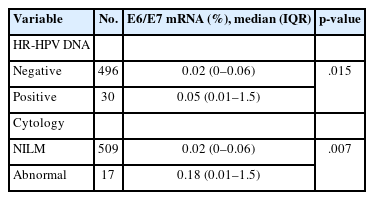
Comparison of E6/E7 mRNA expression according to HPV DNA and cytology status
E6/E7 mRNA detection demonstrated almost perfect concordance with both HR-HPV DNA testing and cytology, as indicated by Gwet’s AC values of 0.94 and 0.96, respectively. However, the PA was relatively low (21.05% with HR-HPV DNA testing and 24.00% with cytology), whereas the NA was 97.04% and 98.15%, respectively (Table 2).
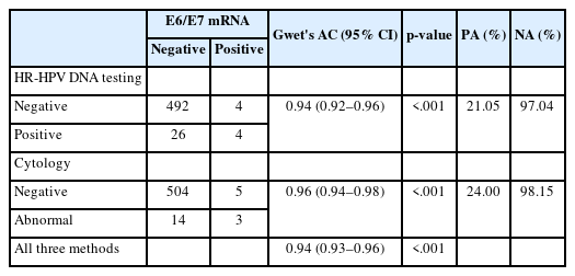
Concordance of E6/E7 mRNA detection with HR-HPV DNA testing and cytological findings
Histopathologic follow-up was available for nine women with cytological abnormalities of LSIL or higher. Histologic examination identified five cases of cervical intraepithelial neoplasia (CIN) 1 and four cases of CIN2. All LSIL cases corresponded to CIN1, whereas all high-grade squamous intraepithelial lesion (HSIL) cases and one atypical squamous cells, cannot exclude high-grade squamous intraepithelial lesion/atypical glandular cells case were confirmed as CIN2 (Table 3).
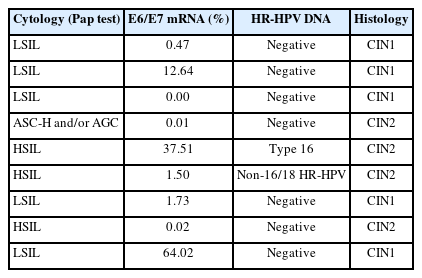
E6/E7 mRNA expression in cases with cytological and histological abnormalities (≥LSIL)
Among the nine participants with histopathologic follow-up, E6/E7 mRNA expression ranged from 0.00% to 64.02%. All five histologically confirmed CIN1 cases were negative for HR-HPV DNA. Two of these cases showed E6/E7 mRNA expression above the 2% positivity threshold (12.64% and 64.02%) (Fig. 1), whereas the other three showed low or undetectable expression. Among the four CIN2 cases, E6/E7 mRNA expression levels were 0.01%, 37.51%, 1.50%, and 0.02%; only one case was E6/E7 mRNA-positive (37.51%) that was observed in an HPV16-positive HSIL case. Overall, six of the nine participants with histologically confirmed CIN1 or CIN2 had E6/E7 mRNA expression below 2% and only one of these six cases tested positive for HR-HPV DNA (Table 3, Fig. 2).
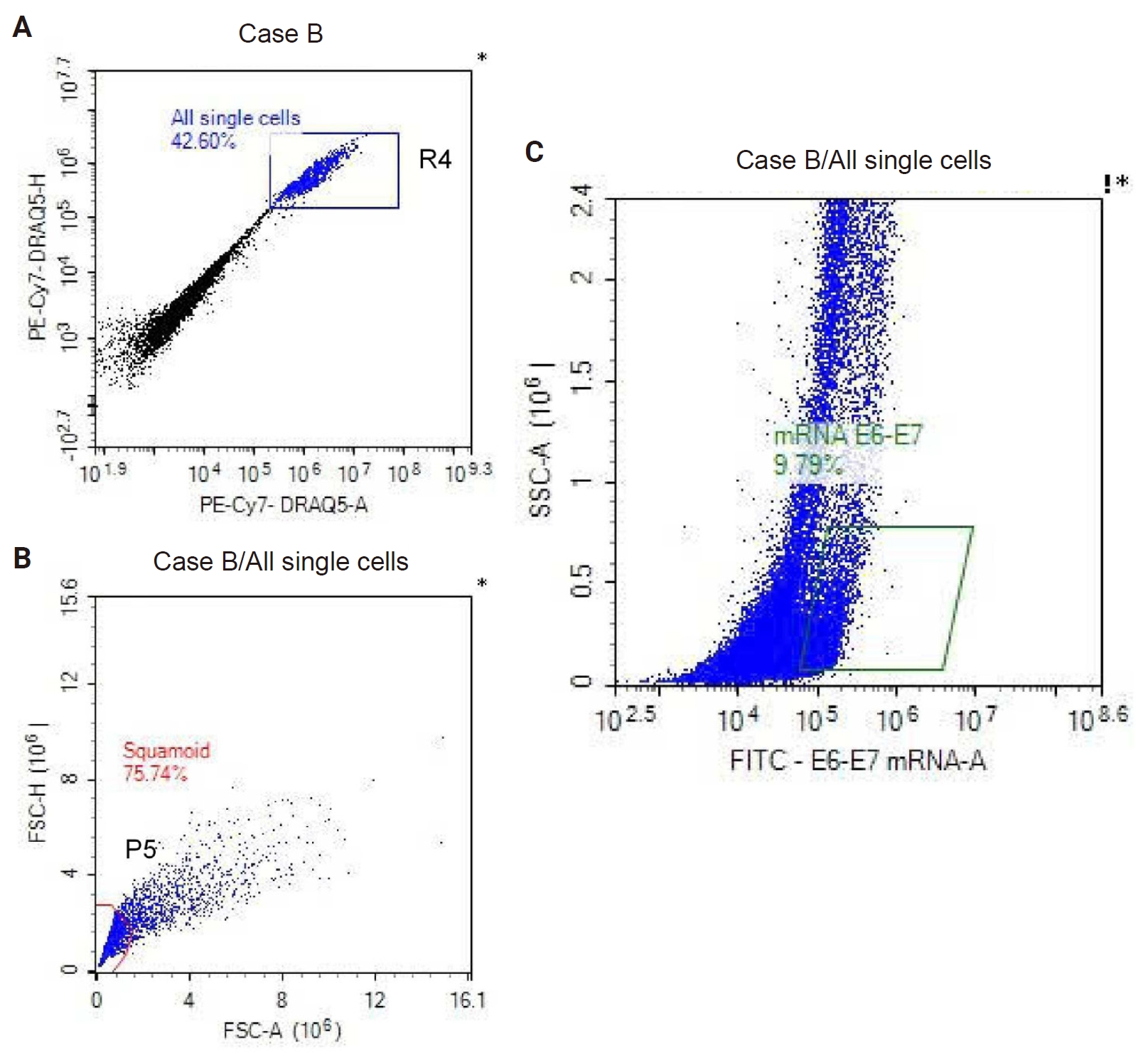
Flow cytometry–based profiling of E6/E7 mRNA expression in an mRNA-positive case. (A) At the R4 region on the PE-Cy7-DRAQ5-H, doublets and debris were excluded and 42.6% of events included in the R4 region were single cells. (B) All these single cells were further gated at the P5 region on the FSC-H/FSC-A dot plot to select cervical epithelial cells, 75.7%. (C) The gated epithelial cell population was subsequently analyzed for E6/E7 mRNA fluorescence. The E6/E7 mRNA-positive population accounted for 9.8% of cells, exceeding the predefined positivity threshold of ≥2% of cells expressing above-threshold E6/E7 mRNA-positive (This is a representative example separate from the eight mRNA-positive cases listed in Table 4).
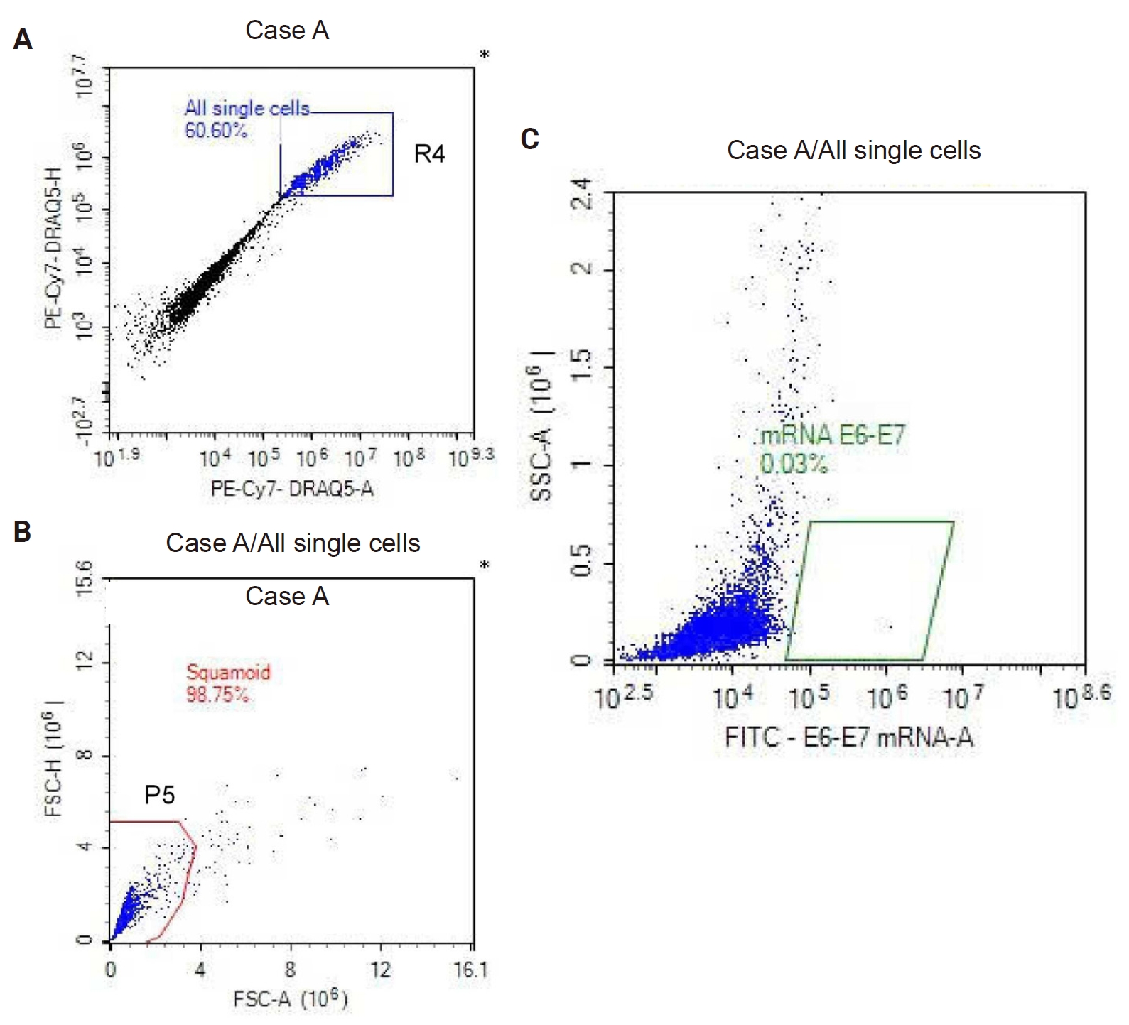
Flow cytometry–based analysis of E6/E7 mRNA expression in an mRNA-negative case. (A) At the R4 region on the PE-Cy7-DRAQ5-H, doublets and debris were excluded and 60.6% of events included in the R4 region were single cells. (B) All these single cells were further gated at the P5 region on the FSC-H/FSC-A dot plot to select cervical epithelial cells, 98.8%. (C) The gated epithelial cell population was subsequently analyzed for E6/E7 mRNA fluorescence. The E6/E7 mRNA–positive population accounted for 0.03% of cells, which was below the predefined positivity threshold of ≥2% of cells expressing above-threshold E6/E7 mRNA signal; therefore, this case was classified as E6/E7 mRNA–negative.
Of the eight E6/E7 mRNA-positive cases, three showed abnormal cytology (LSIL or HSIL), whereas five had negative cytology findings. Four of the eight mRNA-positive cases were also positive for HR-HPV DNA, while the remaining four were negative by HPV DNA testing. Notably, only one E6/E7 mRNA-positive case showed concurrent positivity across all three modalities, namely an HPV16-positive HSIL lesion histologically confirmed as CIN2 (Table 4).
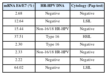
Cytology and HR-HPV results in mRNA E6/E7-positive cases
DISCUSSION
E6/E7 mRNA expression tended to increase in cases with cervical cell abnormalities (p = .007) and in HR-HPV–positive cases (p = .015). These findings align with Kottaridi et al. [9], who also reported that the rate of E6/E7 mRNA positivity increased in parallel with cytological lesion severity. Histopathologic evaluation demonstrated CIN2 lesions in four participants with cytological abnormalities. However, only one showed E6/E7 mRNA expression above the positivity cutoff and this case was an HPV16-positive HSIL/CIN2 case. This isolated finding may suggest a possible association between E6/E7 transcriptional activity and high-grade cervical lesions, particularly in the context of HPV16 infection [4].
In addition, the other three CIN2 lesions exhibited low mRNA expression, and two of them were negative for HR-HPV, suggesting that E6/E7 transcription may vary considerably among lesions of similar histologic grade. In addition, all CIN1 lesions were negative for HR-HPV, and three of five cases expressed E6/E7 mRNA below the positive cutoff. This finding is consistent with existing evidence that, besides HR-HPV, other factors such as chronic Chlamydia trachomatis infection or carcinogens in tobacco smoke can also contribute to cervical epithelial abnormalities, etc. [13,14].
Interestingly, two LSIL cases (histologically CIN1) with high E6/E7 mRNA expression (12.64% and 64.02%) were negative for HPV DNA. While the HR-HPV DNA assay used in this study detects the DNA of 14 HR-HPV genotypes, including HPV16, HPV18, and 12 additional HR-HPV genotypes (31, 33, 35, 39, 45, 51, 52, 56, 58, 59, 66, and 68), the HPV OncoTect 3Dx assay detects E6/E7 mRNA from HR-HPV overall. Therefore, these discordant findings may be related to differences between the two methods in genotype coverage or biological targets. Because histologic follow-up and the number of cases were limited, the clinical significance of these cases remains uncertain.
One HSIL case, positive for HPV16, exhibited very high E6/E7 mRNA expression (37.51%), consistent with the well-established oncogenic potential of this genotype [4]. It is noteworthy that several E6/E7 mRNA–positive cases were negative by cytology and/or HR-HPV DNA testing. Similar results were reported by Kottaridi et al. [9], who found that approximately 10.80% of histologically negative samples tested positive for E6/E7 mRNA. These discordant findings suggest that E6/E7 mRNA testing may provide complementary information to HR-HPV DNA testing and cytology. However, the clinical significance of these discordant cases remains uncertain because longitudinal follow-up and comprehensive histopathologic confirmation were not available.
The present study demonstrated almost perfect agreement between E6/E7 mRNA detection and both HR-HPV DNA testing and cytological findings (Gwet's AC, 0.94 and 0.96, respectively), with very high NA (>97.00%). These findings indicate a high level of concordance between flow cytometry–based E6/E7 mRNA detection and the two established cervical cancer screening methods. However, PA was relatively low (21.05% with HR-HPV DNA testing and 24.00% with cytology), reflecting the small number of E6/E7 mRNA–positive cases in our study population.
Taken together, the findings of this study reinforce the role of E6/E7 mRNA testing as a valuable adjunctive tool in cervical cancer screening, particularly in cases with atypical or discordant cytology/HPV DNA results.
This study has several limitations. Histopathologic confirmation was available only for a limited subset of women with cytological abnormalities, and no CIN3 or invasive carcinoma cases were identified. In addition, the small number of E6/E7 mRNA–positive cases limited the statistical power of the analysis and restricted the evaluation of diagnostic performance. The low prevalence of positive cases may also have affected agreement estimates despite the use of Gwet’s AC. Furthermore, as this was a single-center study, the generalizability of the findings remains limited. Larger prospective studies with comprehensive histopathologic follow-up are needed to further validate the clinical utility of flow cytometric E6/E7 mRNA testing in cervical cancer screening.
Flow-cytometric E6/E7 mRNA detection showed high overall agreement with HR-HPV DNA testing and cervical cytology in this screening cohort. In the small subset with histopathologic follow-up, E6/E7 mRNA expression was heterogeneous among CIN1 and CIN2 lesions, and discordant results were observed. Larger prospective studies with comprehensive histologic and longitudinal follow-up are needed to determine the potential role of this assay in cervical cancer screening and triage.
Notes
Ethics Statement
The study was approved by the Institutional Review Board of Biomedical Research at the University of Medicine and Pharmacy at Ho Chi Minh City (IRB No. 3097/DHYD-HDDD on October 24, 2024) and performed in accordance with the principles of the Declaration of Helsinki. Written informed consent was obtained. The study included retrospectively collected specimens and prospectively enrolled participants after IRB approval.
Availability of Data and Material
The datasets used and analyzed during the current study are available from the corresponding author on reasonable request.
Code Availability
Not applicable.
Author Contributions
Conceptualization: DQN, TDAP. Data curation: DQN, TKH, MNTT, TDAP. Formal analysis: AMTN, LNKN. Investigation: MNTT, AMTN, LNKN, TDAP. Funding acquisition: DQN, TDAP. Methodology: DQN, TKH, AMTN, TDAP. Project administration: DQN, TDAP, AMTN. Resources: DQN, AMTN, TDAP. Software: TNT, TTL. Supervision: TDAP. Validation: DQN, TDAP. Visualization: MNTT, TKH. Writing—original draft: DQN. Writing—review & editing: DQN, AMTN, LNKN, TKH, TDAP. Approval of final manuscript: all authors.
Conflicts of Interest
The authors declare that they have no potential conflicts of interest.
Funding Statement
This study was financially supported by the University of Medicine and Pharmacy at Ho Chi Minh City under Contract No. 121/2024/HĐ-ĐHYD, dated April 17, 2024.