Colorectal intramucosal carcinoma with lymphatic invasion and lymph node metastasis: a case report
Article information

Abstract
Colorectal intramucosal carcinoma (IMC) is considered to carry negligible risk of lymph node metastasis, reflecting the presumed absence of lymphatics within the lamina propria, and current guidelines endorse endoscopic resection alone as curative. We report a 64-year-old man in whom endoscopic submucosal dissection of a lower rectal tumor revealed a well-differentiated IMC with extensive lymphatic invasion confirmed by D2-40 immunostaining. Subsequent laparoscopic ultralow anterior resection showed no residual tumor at the primary site but confirmed metastasis in one of 12 regional lymph nodes. The tumor was microsatellite-stable, epidermal growth factor receptor–positive, and harbored a KRAS codon 13 mutation (G13X). The patient received adjuvant chemoradiotherapy and remained disease-free at 2 years. This case demonstrates that regional lymph node metastasis can occur in colorectal IMC even without conventional adverse histologic features such as poor differentiation or tumor budding, challenging current risk-stratification frameworks and supporting a more cautious approach when lymphatic invasion is identified.
INTRODUCTION
Colorectal intramucosal carcinoma is defined as carcinoma that has invaded the lamina propria or muscularis mucosae but has not penetrated into the submucosa [1]. Despite this invasion into the lamina propria and muscularis mucosae, the World Health Organization classification of digestive system tumors designates such lesions as high-grade dysplasia rather than as intramucosal carcinoma [1]. This classification reflects the well-established anatomical observation that the lamina propria of the colorectum is essentially devoid of lymphatic channels, in contrast to the esophagus and stomach, where lamina propria lymphatics are present and true metastatic potential exists even at the intramucosal stage [2].
Here, we report a case of colorectal intramucosal carcinoma in which definite lymphatic invasion was identified on the endoscopic submucosal dissection (ESD) specimen, prompting additional surgical resection that revealed metastasis in a regional lymph node, despite the absence of residual tumor at the primary site. Given the rarity of both lymphatic invasion and, especially, confirmed nodal metastasis in this setting, we present this case to contribute to the limited evidence in the literature and to raise awareness of this exceptional but clinically significant possibility.
CASE REPORT
A 64-year-old male with no significant past medical history was referred to our institution after a routine health checkup that revealed multiple colonic polyps. Vital signs were within normal limits and he had no gastrointestinal symptoms. Colonoscopy revealed a nodular tumor approximately 2 cm in size at the lower rectum, 5 cm from the anal verge (Fig. 1), and ESD was performed. Additional multiple polyps were scattered throughout the colon and rectum.
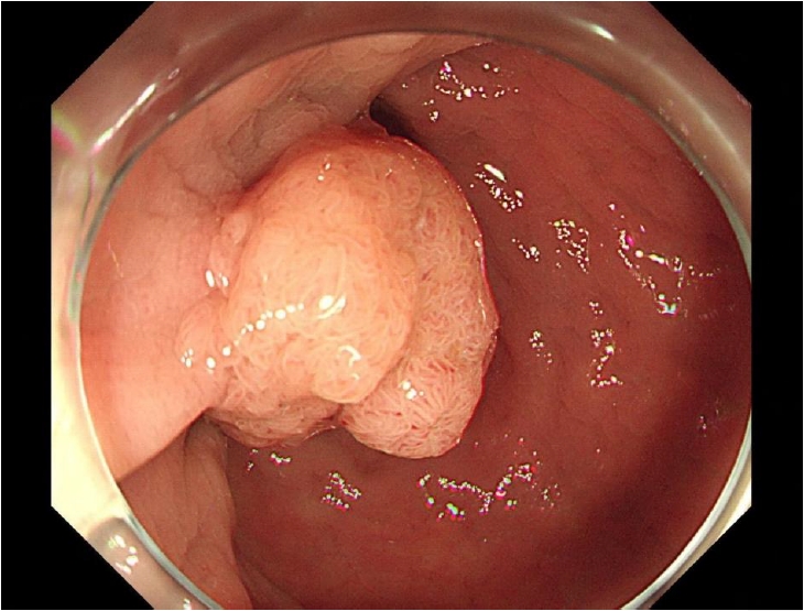
Colonoscopic finding showing a nodular tumor approximately 2 cm in size at the lower rectum, 5 cm from the anal verge.
The ESD specimen was fixed in formalin, serially sectioned at 2-mm intervals, and entirely embedded for slide preparation. Histopathological examination revealed a well-differentiated intramucosal adenocarcinoma arising in a tubulovillous adenoma, measuring 1.6 × 1.0 cm including the adenomatous component (Fig. 2). The tumor cells showed marked cytologic atypia with round, bizarre nuclei and plump cytoplasm, without definite tumor budding. The carcinoma invaded up to the muscularis mucosae without extension into the submucosa, which was confirmed by desmin immunohistochemical staining delineating the muscularis mucosae layer (Fig. 3). Prominent and unequivocal lymphatic tumor invasion was identified and confirmed by D2-40 immunohistochemical staining (Figs. 4, 5). The resection margin was clear.
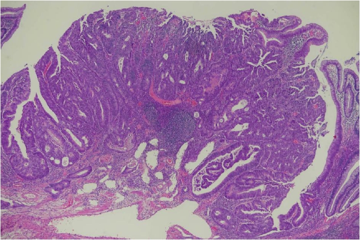
Low-power photomicrograph of the endoscopic submucosal dissection specimen showing a well-differentiated intramucosal adenocarcinoma arising within a tubulovillous adenoma.
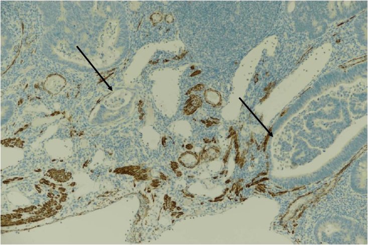
Desmin immunohistochemical staining delineates the smooth muscle fibers of the muscularis mucosae, confirming that tumor invasion is confined within this layer without submucosal extension (desmin). Arrows indicate carcinoma invading muscularis mucosa.
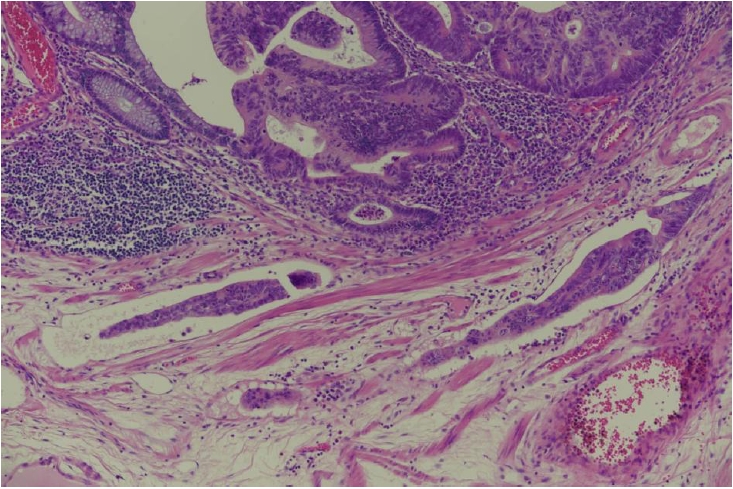
Tumor cell clusters are identified within dilated vascular spaces in the muscularis mucosae, consistent with lymphatic invasion. The tumor cells show eosinophilic cytoplasm, round nuclei, and peritumoral inflammation.
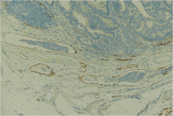
D2-40 immunohistochemical staining highlights the endothelial lining of the involved vascular spaces, confirming true lymphatic invasion and excluding retraction artifact (D2-40).
Given the presence of lymphatic invasion, the patient subsequently underwent laparoscopic ultralow anterior resection. Histopathological examination of the surgical specimen revealed no residual tumor at the ESD site, without venous or perineural invasion. Of the 12 regional lymph nodes examined, metastatic carcinoma was identified in 1 lymph node (Figs. 6, 7).
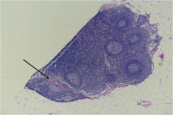
Low-power view of a regional lymph node showing a discrete focus of metastatic adenocarcinoma (arrow).
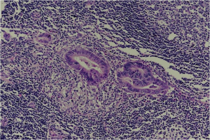
High-power view of the metastatic focus within the lymph node, demonstrating glandular architecture consistent with the primary colorectal adenocarcinoma.
KRAS mutation analysis, performed by real-time polymerase chain reaction (PCR) with peptide nucleic acid (PNA) clamping (PNAClamp KRAS Mutation Detection Kit ver. 4, Panagene Inc., Daejeon, Korea) following DNA extraction with the QIAamp DSP DNA FFPE Tissue Kit (Qiagen, Hilden, Germany), identified a codon 13 mutation (G13X), while NRAS mutation analysis was negative. Microsatellite instability testing, performed by real-time PCR (U-TOP MSI Detection Kit, Seasun Biomaterials Inc., Daejeon, Korea) using five microsatellite markers (BAT25, BAT26, NR21, NR24, and NR27) following DNA extraction with the QIAamp DSP DNA FFPE Tissue Kit (Qiagen), showed a microsatellite-stable status. Immunohistochemical staining was performed using an automated immunostainer (BenchMark ULTRA, Ventana Medical Systems, Tucson, AZ, USA) with the OptiView DAB IHC Detection Kit (Ventana Medical Systems), using antibodies against epidermal growth factor receptor (EGFR; clone SP84, Cell Marque, Rocklin, CA, USA) and pan–tropomyosin receptor kinase (pan-TRK; clone EPR17341, Ventana Medical Systems). The tumor cells were positive for EGFR and negative for pan-TRK.
Following surgery, the patient received postoperative radiotherapy and adjuvant chemotherapy with capecitabine. There has been no evidence of local recurrence or distant metastasis for 2 years.
DISCUSSION
The colorectal mucosa is known to have few, if any, lymphatic vessels within the lamina propria [3]. However, subsequent studies using the lymphatic endothelial marker D2-40 have demonstrated that lymphatic vessels can indeed be identified within the colonic lamina propria under pathologic conditions [4,5]. Kenney and Jain found that such lymphatics were largely absent in normal colonic mucosa but were frequently present in inflamed mucosa, hyperplastic and adenomatous polyps, and especially in polyps containing intramucosal carcinoma, suggesting that this lymphangiogenesis is induced by the underlying inflammatory or neoplastic process rather than representing a baseline feature of the mucosa [5]. These findings indicate that lymphatic vessels are not constitutively present in the normal colonic mucosa but can be induced by underlying inflammatory or neoplastic processes—an "aberrant" lymphangiogenesis that may explain how lymphatic invasion, and even lymph node metastasis, can occur in lesions histologically confined to the mucosa [6].
Lymphatic invasion in colorectal intramucosal carcinoma has rarely been reported [7,8]. Hashimoto et al. [7] reported the clinicopathologic characteristics of nine cases of intramucosal colorectal carcinoma with lymphovascular invasion, finding that high-grade tumor budding and an "eosinophilic cytoplasm and round nuclei with inflammatory reaction" (ERI) pattern were characteristic of these cases. In our case, the well-differentiated adenocarcinoma showed somewhat eosinophilic cytoplasm and round nuclei, but no evidence of tumor budding. Whether the ERI pattern truly represents a characteristic feature of lymphatic invasion in colorectal intramucosal carcinoma remains to be clarified as more cases accumulate.
Vink-Borger et al. [9] reviewed 14 strictly defined cases of true colorectal intramucosal carcinoma, applying rigorous histologic criteria to exclude high-grade dysplasia and invasive carcinoma. The study reported an extremely low population incidence (0.01%), a low risk of lymph node involvement, and generally favorable patient outcomes—consistent with the rarity of lymphatic invasion and nodal metastasis observed across the literature. Notably, however, the cohort also showed a strong association with hereditary cancer syndromes, particularly Lynch syndrome, with deficient mismatch repair (dMMR) present in up to 50% of cases, nearly five times the rate seen in the general colorectal cancer population.
The molecular profile of our tumor differed from this pattern. The tumor was microsatellite-stable, EGFR-positive by immunohistochemistry, and harbored a KRAS codon 13 mutation (G13X), with negative pan-TRK and NRAS testing. Unlike the dMMR-enriched cohort described by Vink-Borger et al. [9], our patient had multiple colonic polyps that did not meet the criteria for a polyposis syndrome, and the microsatellite-stable status suggests a sporadic rather than Lynch syndrome–associated pathway.
KRAS mutation has been associated with adverse histopathological features such as poor differentiation, lymph node metastasis, and perineural invasion [10-12]; however, these associations have been reported mainly in T1 or more advanced tumors rather than in strictly intramucosal carcinoma. Accordingly, the KRAS-positive status in this case should be regarded as a possible contributing factor rather than a definitive explanation for the observed lymphatic invasion and nodal metastasis. To our knowledge, no prior studies have specifically examined the relationship between molecular alterations and lymphatic invasion in intramucosal carcinoma, and this remains an area for future investigation.
Lymph node metastasis from colorectal intramucosal carcinoma is exceedingly rare [13-15]. Across the largest and most recent cohort studies, the reported rate is essentially 0% [7,16]. In our case, definite lymphatic invasion was accompanied by regional lymph node metastasis, and the patient was subsequently treated with adjuvant chemoradiotherapy.
Global guidelines classify intramucosal carcinoma as high-grade dysplasia with virtually zero lymph node metastasis risk, treating it definitively with complete endoscopic resection. Endoscopic resection is considered curative when margins are clear and there is no lymphovascular or submucosal invasion, according to the National Comprehensive Cancer Network and European Society for Medical Oncology criteria [17,18].
From a clinical standpoint, this case highlights a gap in current management guidance. The European Society of Gastrointestinal Endoscopy guidelines, developed for T1 carcinomas with submucosal invasion, treat lymphovascular invasion as grounds for additional treatment—but no comparable framework exists for carcinomas confined to the mucosa [19]. Once lymphatic invasion is identified in such a lesion, it remains unclear whether the appropriate response is close surveillance, oncologic surgery, or another approach. Based on this experience, we suggest that additional oncologic resection with lymph node evaluation is advisable over surveillance alone when lymphatic invasion is confirmed in intramucosal carcinoma, pending further data.
This report has several limitations. As a single case, it cannot establish how often lymphatic invasion in colorectal intramucosal carcinoma progresses to nodal metastasis, nor can it identify which histologic or molecular features reliably predict this progression. Follow-up in a single patient is also insufficient to confirm durable oncologic control, although the absence of recurrence or distant metastasis at 2 years after surgical resection and adjuvant therapy is reassuring.
In conclusion, we report a case of colorectal intramucosal carcinoma with D2-40–confirmed lymphatic invasion and subsequent regional lymph node metastasis, despite the absence of residual tumor at the primary site. This case challenges the assumption that lesions confined to the mucosa carry no metastatic potential, and supports a more cautious approach to management when lymphatic invasion is identified, even in intramucosal disease.
Notes
Ethics Statement
This case was reviewed and the requirement for informed consent was waived by the Institutional Review Board of Inha University Hospital (IRB No. 2026-07-020), in accordance with the 1964 Helsinki Declaration and its later amendments.
Availability of Data and Material
Data sharing not applicable to this article as no datasets were generated or analyzed during the study.
Code Availability
Not applicable.
Author Contributions
Conceptualization: JMK. Data curation: LK, JYH. Investigation: JS, SKC. Methodology: SJC. Resources: JS, SKC. Supervision: JMK. Writing—original draft: JMK. Writing—review & editing: LK, JYH, SJC, JS, SKC. Approval of final manuscript: all authors.
Conflicts of Interest
The authors declare that they have no potential conflicts of interest.
Funding Statement
This work was supported by Inha University.