Primary hepatic extranodal marginal zone lymphoma of mucosa-associated lymphoid tissue
Article information

Abstract
Extranodal marginal zone lymphoma of mucosa-associated lymphoid tissue (MALT lymphoma), is one of the specific type of low-grade B-cell lymphoma not infrequently found worldwide. It typically involves mucosal sites such as stomach and conjunctiva; however, primary hepatic MALT lymphoma has been extremely rarely reported. We describe a case of hepatic MALT lymphoma in a 70-year-old male patient who underwent left hepatectomy due to the incidentally detected liver masses at a medical checkup. The resected specimen revealed multinodular masses consisting of small-to-intermediate-sized lymphoid cells with serpentine pattern and focal lymphoepithelial lesions. The tumor cells were diffusely positive for CD20 and Bcl-2 but negative for CD3, CD10, CD5, CD23, CD43, and cyclinD1. The Ki-67 labeling index was 10% and immunoglobulin heavy chain gene rearrangement study confirmed monoclonal proliferation. In this paper, we discuss several unique clinicopathologic characteristics which will be helpful to the differential diagnosis of hepatic MALT lymphoma.
Extranodal marginal zone lymphoma of mucosa-associated lymphoid tissue (MALT) is one of the specific type of low-grade B-cell lymphoma not infrequently found worldwide. MALT lymphoma represents 7%–8% of all non-Hodgkin’s lymphomas in Western countries [1]. In Korea, MALT lymphomas represent 19.0% of non-Hodgkin lymphomas and over half (59.8%) occur in the stomach. Other sites of occurrence include the lung, salivary gland, ocular adnexa, skin, and thyroid [2]. However, lymphoma in the liver as the primary site is very rare. Primary hepatic lymphoma (PHL) comprises 0.016% of non-Hodgkin’s lymphoma [3] and MALT lymphoma comprises 2%–4% of PHL [4]. Since it is a rare disease with limited literature, there is no consensus on treatment modalities [5,6]. Here, we report a case of primary hepatic MALT lymphoma.
CASE REPORT
A 70-year-old male patient was referred for liver masses on ultrasonography from a medical checkup at another hospital. He had a history of intracerebral hemorrhage, and coronary stent insertion due to myocardial infarction. A physical examination showed no abnormalities. The laboratory test results were within normal limits. The level of aspartate transaminase was 27 IU/L, alanine transaminase was 24 IU/L, total bilirubin was 0.9 mg/dL, alkaline phosphatase was 96 IU/L, and γ glutamyl transferase was 34 IU/L. The patient was seronegative for hepatitis B and C and tumor markers (α-fetoprotein and protein induced by vitamin K antagonist II) were within normal limits. He was also seronegative for human immunodeficiency virus and was negative for antinuclear antibody. The abdomen ultrasonography showed mild fatty liver. Esophagogastroduodenoscopy showed atrophic gastritis and no other MALT lesion was present. Further imaging studies including computed tomography (CT), magnetic resonance imaging (MRI), and 18Ffluorodeoxyglucose (FDG)-positron emission tomography (PET)/CT were performed. The MRI revealed two arterial enhancing masses measuring 3.6 cm and 3.3 cm in the left lateral segment of the liver, suggesting hepatocellular carcinoma (HCC) (Fig. 1A). The PET/CT revealed the two masses in the left lateral segment of the liver to be hypermetabolic (maximum standardized uptake value of 4.4) (Fig. 1B). No other lymphadenopathy was present. He underwent left hepatectomy.
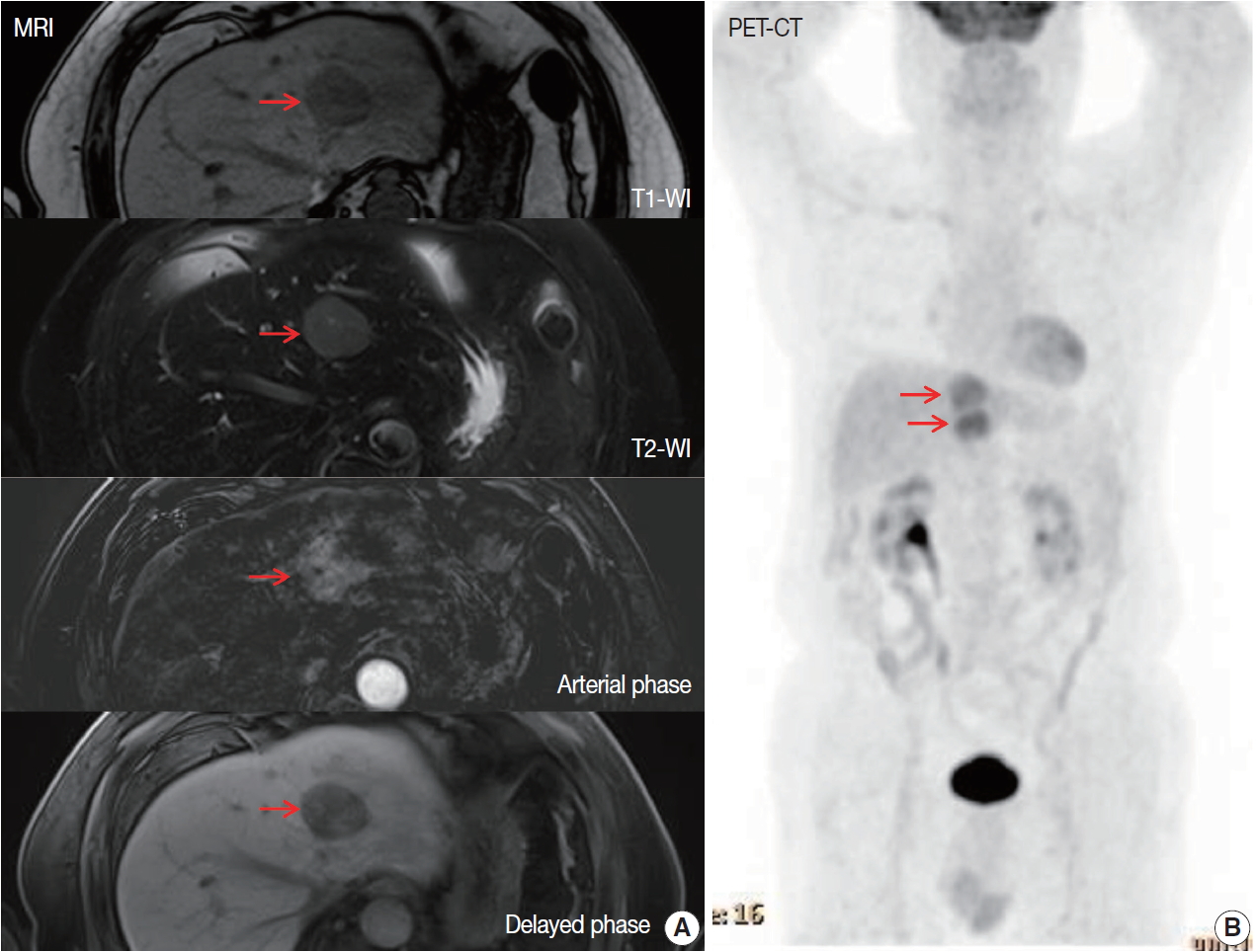
(A) The magnetic resonance imaging shows a hypointense in T1, and T2, and arterial enhancing lesion in left lateral segment (arrows). (B) The positron emission tomography/computed tomography shows two hypermetabolic masses in left lateral segment (maximal standardized uptake value, 4.4). T1-WI, T1 weighted image; T2-WI, T2 weighted image.
The liver specimen showed two masses measuring 4.8 × 4.2 × 3.3 cm and 4.8 × 3.8 × 2.3 cm that were well-demarcated and grayish tan in color in segment 2 (Fig. 2A). On low-power magnification, nodular and serpentine infiltration of lymphocytes was seen (Fig. 2B). On high-power magnification, small-to-intermediate-sized lymphoid cells were present (Fig. 2C). Lymphoepithelial lesions were present in the bile ducts (Fig. 2D), which were highlighted by positive immunoreactivity for cytokeratin 7 by immunohistochemistry (IHC) (Fig. 2E). In other IHC studies, CD20 and Bcl-2 were diffusely and strongly positive, and the tumor cells were negative for CD3, CD10, CD5, CD23, CD43, and cyclinD1 (Fig. 3A–C). The Ki-67 index was about 10%. Immunoglobulin heavy chain (IgH) gene rearrangement study revealed monoclonality of the lymphoid lesion, and all these results were compatible with a diagnosis of MALT lymphoma. The background liver outside the nodules showed portal-to-periportal lymphocytic infiltration (Fig. 3D), however, there was no evidence of autoimmune hepatitis, or primary biliary cirrhosis, or non-alcoholic steatohepatitis. Systemic studies including bone marrow biopsy showed no additional lesions. He was diagnosed with Ann Arbor stage IIE and close follow-up without additional therapy was recommended. After 8 months from the surgery, the patient showed no recurrent disease in the CT scan.
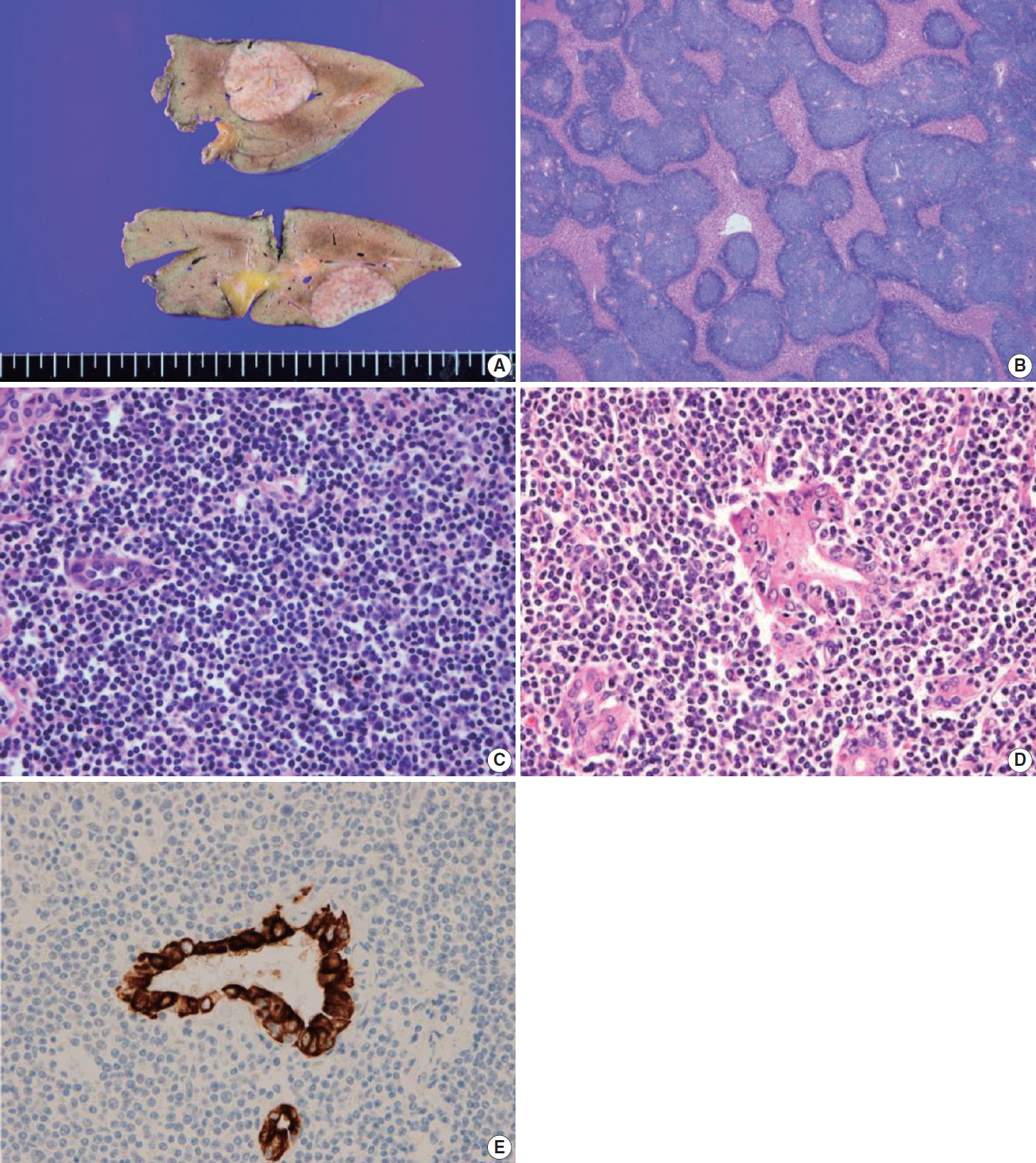
(A) Gross image of the liver shows well-demarcated and grayish tan masses. (B) Low power view of the lesion demonstrates serpentine infiltrations of lymphoid cells. (C) High power view of the lesion demonstrates small-to-intermediated-size lymphoid cell infiltration. (D, E) Lymphoepithelial lesion is present, which is revealed by cytokeratin 7 immunohistochemical stain.
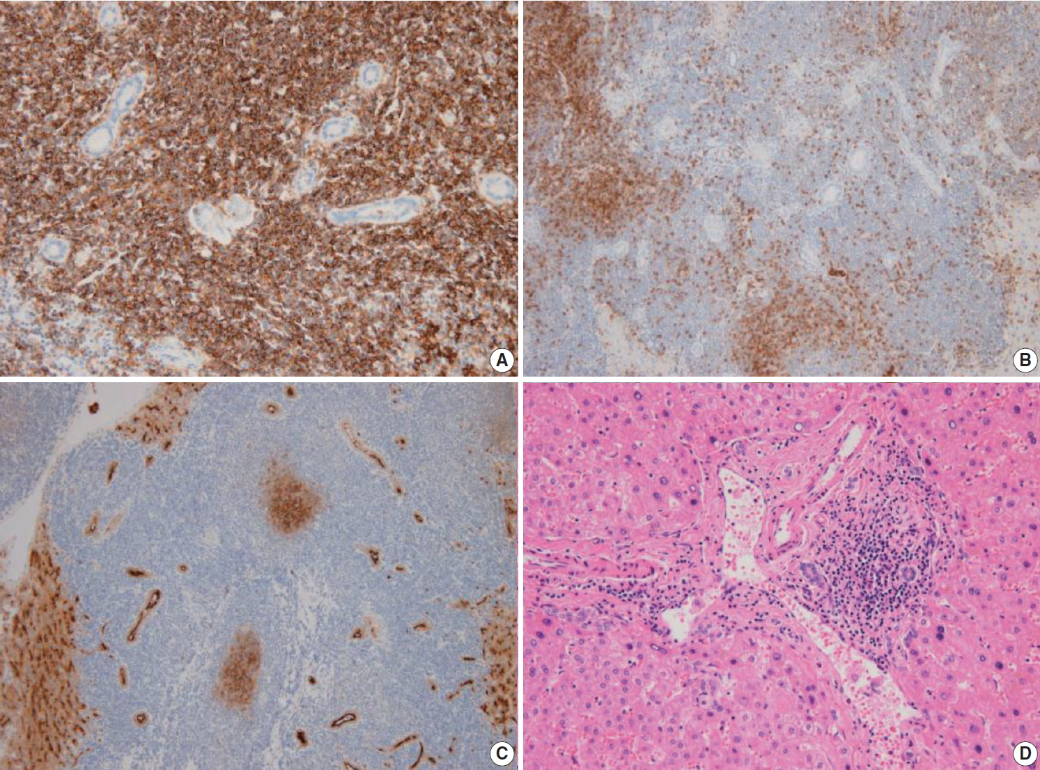
The lymphoid cells are positive for CD20 (A), and negative for CD3 (B) and CD10 (C). (D) The non-neoplastic liver shows portal to periportal lymphocytic infiltration.
DISCUSSION
Although the incidence of primary hepatic MALT lymphoma is extremely rare, some unique clinical characteristics were reported in the literatures (Table 1) [6-15]. Most of primary hepatic MALT lymphoma cases occurred in elderly people and were presented with incidentally detected liver masses without specific symptoms [6,7]. These features were consistent with the present case.
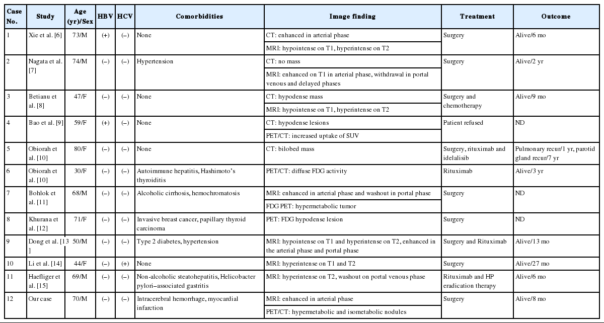
Reported cases of primary hepatic MALT lymphoma
Chronic antigenic stimulation such as longstanding inflammation or infection has been suggested as an important etiologic factor of MALT lymphoma. Helicobacter pylori has been proved to be the cause of gastric MALT lymphoma and the suggested infectious agents for other sites are Borrelia burdorferi for skin, Chlamydophila psittaci for ocular adnexa, Campylobacter jejuni for small intestine, and Achromobacter xylosoxidans for lung. As for hepatic MALT lymphoma, hepatitis C viral infection, autoimmune diseases such as Sjogren’s disease or Hashimoto’s thyroiditis have been reported to have increased the risk of MALT lymphoma [16,17]. Another study proposed that 45% of patients with MALT lymphoma had no concomitant liver diseases [6]. Our patient had no suggested predisposing conditions listed above. The pathogenesis of hepatic MALT lymphoma is still unclear and should be explored in a large-scale study.
In this case, the patient was initially misdiagnosed with HCC by CT and MRI studies. Betianu et al. [8] states that there are no characteristic radiological findings of PHL. After contrast administration, more than 50% of PHLs showed no enhancement, 30% showed patchy enhancement and 15% showed a ring enhancement. On MRI, PHL is hypo- or isointense on T1 and hyperintense on T2 [8]. Thus, when suspected malignant liver masses are seen in imaging studies without elevation of tumor markers, lymphoma should be a differential diagnosis and liver biopsy could be an option. Several articles focus on 18F-FDG PET/CT as a diagnostic modality as for other lymphomas. Bao et al. [9] suggest that 18F-FDG PET/CT is particularly helpful in distinguishing the initial stages of MALT lymphoma, although pathological evidence is needed for definitive diagnosis. It can assist in staging, evaluating treatment responses, and monitoring relapse and recurrence [9]. Our patient had a slightly hypermetabolic mass, considering its indolent nature. The standardized uptake value of primary hepatic MALT lymphoma remains to be validated with more evidence.
Microscopically, in low-power magnification, nodular and serpentine infiltrations of lymphoid cells from the portal area were present. MALT lymphomas are known to predominantly involve the portal fields of the liver, and lymphoepithelial lesions of the bile ducts are known to be typical accompanying findings [18]. The differential diagnoses are HCC, cholangiocellular carcinoma, low-grade B-cell lymphoma including follicular lymphoma and small lymphocytic lymphoma, hepatosplenic T-cell lymphoma, pseudolymphoma, and IgG4-related liver disease. IHC with CD5, CD10, CD23, CD43, and cyclinD1 is critically important to further classify low-grade B-cell lymphomas. Hepatic pseudolymphoma is another rare lesion often associated with inflammatory or autoimmune diseases and shows features similar to malignant lesions in imaging. Its histologic findings resemble low-grade lymphoma, particularly extranodal marginal zone lymphoma [19]. It can be distinguished from extranodal marginal zone lymphoma by histology, and IHC studies showing reactive patterns and polyclonality in an IgH gene rearrangement study. The diagnostic features of IgG4-related disease include elevated serum IgG4 concentrations, and histologic features include dense lymphoplasmacytic inflammatory infiltrate with increased numbers of IgG4+ plasma cells, often increased eosinophils, a storiform pattern of fibrosis, and obliterative vasculitis [20]. The histologic findings along with results from IHC study and IgH gene rearrangement study seem to be critical in the differential diagnosis.
H.pylori eradication is a well-established treatment for gastric MALT lymphoma. However, there is no consensus on the treatment for MALT lymphoma in other sites. Expectant observation, surgery, radiotherapy, chemotherapy, and immunotherapy are generally performed, considering site, disease stage, and performance status. For localized diseases, radiation therapy is often a preferred option and surgery can be considered. For advanced stages, observation with monitoring was often proven to be adequate, but when treatment is required, chemotherapy with rituximab and enrollment in controlled clinical trials is suggested [5]. After discussion, our patient was placed under observation. Tumor recurrence has been reported in the literature, with the lung reported as the most common site [10].
Primary hepatic MALT lymphoma is a very rare disease. The clinical features are nonspecific and the image findings variable. Histology, IHC studies and an IgH gene rearrangement study are of great importance in accurate diagnosis. The disease characteristics should be disclosed further with more data.
Notes
Ethics Statement
This study was approved by the Institutional Review Board of Ulsan University Hospital with a waiver of informed consent (IRB No. 2019-12-002).
Author contributions
Conceptualization: HJC (Hee Jeong Cha), KK.
Investigation: HJC (Hee Jeong Cha), KK, SC.
Supervision: HJC (Hee Jeong Cha).
Visualization: HJC (Hee Jeong Cha), KK, MJS.
Writing—original draft: HJC (Hee Jeong Cha), KK, SC.
Writing—review & editing: JHS, YMK, HJC (Hye Jeong Choi), HJC (Hee Jeong Cha), KK, MK, MJS, JHK, SC.
Conflicts of Interest
The authors declare that they have no potential conflicts of interest.
Funding
No funding to declare.