E-submission
Articles
- Page Path
- HOME > J Pathol Transl Med > Volume 48(1); 2014 > Article
-
Brief Case Report
A Rare Case of Pulmonary Papillary Adenoma in Old Aged Woman: A Brief Case Report - In Ho Choi, Joungho Han, Jung Won Moon1, Yong Soo Choi2, Kyung-Jong Lee3
-
Korean Journal of Pathology 2014;48(1):66-68.
DOI: https://doi.org/10.4132/KoreanJPathol.2014.48.1.66
Published online: February 25, 2014
Department of Pathology, Samsung Medical Center, Sungkyunkwan University School of Medicine, Seoul, Korea.
1Department of Radiology, Samsung Medical Center, Sungkyunkwan University School of Medicine, Seoul, Korea.
2Department of Thoracic Surgery, Samsung Medical Center, Sungkyunkwan University School of Medicine, Seoul, Korea.
3Department of Medicine, Samsung Medical Center, Sungkyunkwan University School of Medicine, Seoul, Korea.
- Corresponding Author: Joungho Han, M.D. Department of Pathology, Samsung Medical Center, Sungkyunkwan University School of Medicine, 81 Irwon-ro, Gangnam-gu, Seoul 135-710, Korea. Tel: +82-2-3410-2800, Fax: +82-2-3410-0025, 'hanjho@skku.edu'
© 2014 The Korean Society of Pathologists/The Korean Society for Cytopathology
This is an Open Access article distributed under the terms of the Creative Commons Attribution Non-Commercial License (http://creativecommons.org/licenses/by-nc/3.0/) which permits unrestricted non-commercial use, distribution, and reproduction in any medium, provided the original work is properly cited.
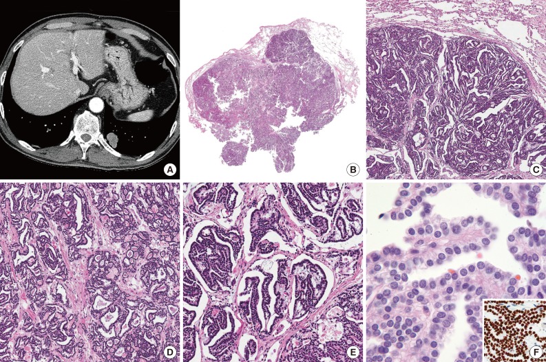
- A 68-year-old female, who was undergoing preoperative evaluation for known rectal cancer, received percutaneous needle biopsy for a pulmonary nodule (Fig. 1A) and endobronchial sono-guided biopsy for enlarged mediastinal lymph nodes, which showed no evidence of metastasis. However, the core of the pulmonary nodule was composed of bland papillae-like structures lined with a single layer of uniform cuboidal cells, suspicious for carcinoid or low-grade epithelial neoplasm. On consideration of double primary pulmonary neoplasm, she received combined laparoscopic low anterior resection of the rectum and wedge resection of the left lobe of the lung.
- The resected specimen of lung contained a 2.5×1.5-cm-sized, peripherally located mass with a granular cut surface. At low power view, it is well demarcated without necrosis or hemorrhage (Fig. 1B). Microscopically, it showed dominant papillary architecture and focal microcystic area (Fig. 1C, D). The papillae contained prominent or delicate fibrovascular cores and were lined by monotonous cuboidal cells (Fig. 1E). The epithelial cells had moderate or small amount of cytoplasm and round, centrally located nuclei with inconspicuous nucleoli, and no nuclear atypia or mitosis was found (Fig. 1F). The chromatin was relatively uniformly distributed and finely granular, suspicious for neuroendocrine tumor. The microcystic area were lined with same epithelial cells and filled with amorphous eosinophilic material, similar to colloid or amyloid.
- For the differential diagnoses with papillary adenoma of lung, metastatic follicular or medullary carcinoma of thyroid gland and less likely metastatic adenocarcinoma of rectum, immunohistochemical staining was performed. The tumor cells showed negativity for chromogranin (1:400, DAK-A3, Dako, Glostrup, Denmark), thyroglobulin (1:500, DAK-Tg6, Dako), calcitonin (1:4,000, Dako) and CDX-2 (1:50, AMT28, Novocastra, Newcastle upon Tyne, UK) and positivity for thyroid transcription factor-1 (1:100, 8G7G3/1, Dako) (Fig. 1F, inset). The eosinophilic material was negative by Congo-red stain and polarizing microscopy.
- The patient was confirmed to have primary papillary adenoma of the lung and adenocarcinoma of the rectum (pT3N0Mx), and was discharged without any complications.
CASE REPORT
- Papillary adenoma is a rare pulmonary neoplasm for which 23 cases (range of age, 7 to 66 years in English literature) have been reported to date. We add a case of papillary adenoma that occurred in a 68-year-old woman.
- Although the present case seemed to be microscopically simple and uneventful, the rarity of the tumor and its uncommon finding at intraoperative consultation made it possible to be mistaken as malignancy, as we suggested primary or metastatic adenocarcinoma on frozen section due to solid area with mild atypia. Nakano et al.2 reviewed previously reported 16 cases and their one case of pulmonary papillary adenoma, which four cases of them had description of intraoperative consultation by the frozen section or imprint cytology. Only one case was diagnosed as papillary adenoma on frozen section, while the possibilities of malignancy were not excluded in other three cases. They also concluded that one of the diagnostic clues might be the macroscopic findings, in which the fresh tumor mass had a granular cut surface that became gradually obscure in shape and leaked out from the stump during manipulation of the frozen section.
- The nuclear feature and monotonous appearance were sufficient to suggest carcinoid tumor as the first impression, after which repeated chromogranin stains for the biopsied and operated specimens were performed. Although carcinoid tumor also can show a papillary or follicular growth pattern, most of them grow in an organoid nesting or trabecular arrangement, and single-layered tumor cells regardless of growth pattern may be unusual in carcinoid tumor. On the review of cytological features of previously reported cases, two cases reported by Hegg et al.5 were described similar to present case, but no immunohistochemical study was performed. Although two cases reported by Dessy et al.6 included with chromogranin A and synaptophysin analysis and revealed negative results, those histologic findings were very different from our case, having no impression of carcinoid tumor. One case that was suspected to papillary adenoma with carcinoid-like feature was reported by Yamamoto et al.,7 in which peripherally-located papillary adenoma were histologically very similar to our case and showed negativity for chromogranin staining.
- Metastatic thyroid cancer was also considered due to papillary or cyst-like structures lined by monotonous cuboidal cells resembling those of thyroid follicles and colloid-like material. However, the nuclear features are different from those of papillary thyroid carcinoma, and the results of immunohistochemical and Congo-red staining can exclude the possibilities of metastatic follicular or medullary carcinoma.
- We previously experienced cases of fetal adenocarcinoma of the lung that showed irregularly-shaped tubules, papillary structure and a minute rosette-like growth pattern. However, they also showed pseudostratified lining epithelium, unlike the uniform single-layered epithelium of the present case.
- Of benign pulmonary neoplasm, alveolar adenoma should be included in the differential diagnosis due to shared histologic findings with papillary adenoma. Alveolar adenoma can show a papillary structure, but it mainly consists of variable-sized cysts filled with proteinaceous material, variably thickened septa and a centrally located large cyst. Even though this case showed multiple small cysts, the predominant papillary structure and lack of spindle and inflammatory cells suggest papillary adenoma rather than alveolar adenoma.4 The peripheral type of glandular papilloma also can be considered due to papillary fronds, but its epithelial lining consists of a stratified columnar or cuboidal epithelium and mucous cells with varying proportion.8 This is distinct from our case that consisted of overall uniform cuboidal cells having centrally located bland nuclei.
- Papillary adenoma of the lung is not often encountered, considering only one case report of pulmonary papillary adenoma in Korea.3 However, if we encounter such a case, it will be a difficult situation that asks pathologists many considerations, especially when the tumor shares carcinoid-like features or complicated clinical situation. Herein, we report a case of papillary adenoma of the lung, mimicking carcinoid tumor and neoplasm of thyroid gland.
DISCUSSION
- 1. Spencer H, Dail DH, Arneaud J. Non-invasive bronchial epithelial papillary tumors. Cancer 1980; 45: 1486–1497. PMID: 7357529. ArticlePubMed
- 2. Nakano T, Yokose T, Hasegawa C, et al. Papillary adenoma of the lung with a peculiar raw macroscopic feature. Pathol Int 2011; 61: 475–480. PMID: 21790862. ArticlePubMed
- 3. Sohn ST, Jeong TY, Lee WM, et al. Papillary adenoma of the lung with pulmonary sequestration: a case report. Korean J Thorac Cardiovasc Surg 1997; 30: 1262–1266.
- 4. Travis W, Brambilla E, Müller-Hermelink K, Harris CC. World Health Organization classification of tumours: pathology and genetics of tumours of the lung, pleura, thymus and heart. Lyon: IARC Press, 2004; 82–84.
- 5. Hegg CA, Flint A, Singh G. Papillary adenoma of the lung. Am J Clin Pathol 1992; 97: 393–397. PMID: 1543163. ArticlePubMed
- 6. Dessy E, Braidotti P, Del Curto B, et al. Peripheral papillary tumor of type-II pneumocytes: a rare neoplasm of undetermined malignant potential. Virchows Arch 2000; 436: 289–295. PMID: 10782889. ArticlePubMed
- 7. Yamamoto T, Horiguchi H, Shibagaki T, Kamma H, Ogata T, Mitsui K. Encapsulated type II pneumocyte adenoma: a case report and review of the literature. Respiration 1993; 60: 373–377. PMID: 8290804. ArticlePubMed
- 8. Aida S, Ohara I, Shimazaki H, et al. Solitary peripheral ciliated glandular papillomas of the lung: a report of 3 cases. Am J Surg Pathol 2008; 32: 1489–1494. PMID: 18708941. ArticlePubMed
References

Figure & Data
References
Citations
- Pulmonary papillary adenoma with malignant potential: a case report and literature review
Ping Liu, Junjian Feng, Min Yang, Jingqiu Chen, Luyao Fu, Junxu Lu
Diagnostic Pathology.2022;[Epub] CrossRef - Central papillary adenoma of the lung diagnosed in a bronchoscopy-guided FNA: Cytological and histological characterization of this rare entity
Iñigo Gorostiaga, Adriano Martinez-Aracil, Blanca Catón, Alvaro Perez-Rodriguez
Revista Española de Patología.2021; 54(3): 206. CrossRef - Retrospective study of clinical and pathologic features of pulmonary papillary adenoma
Pengcheng Zhou, Wei Yu, Li Wang, Qianming Xia, Keling Chen
Medicine.2020; 99(44): e23066. CrossRef - Pulmonary papillary adenoma presenting in central portion: a case report
Xu-Yong Lin, Qiang Han, En-Hua Wang, Yong Zhang
Diagnostic Pathology.2015;[Epub] CrossRef
 PubReader
 Cite this Article