E-submission
Articles
- Page Path
- HOME > J Pathol Transl Med > Volume 46(5); 2012 > Article
-
Case Report
Castleman's Disease of the Renal Sinus Presenting as a Urothelial Malignancy: A Brief Case Report - Se Min Jang, Hulin Han, Ki-Seok Jang, Young Jin Jun, Tchun Yong Lee1, Seung Sam Paik
-
Korean Journal of Pathology 2012;46(5):503-506.
DOI: https://doi.org/10.4132/KoreanJPathol.2012.46.5.503
Published online: October 25, 2012
Department of Pathology, Hanyang University College of Medicine, Seoul, Korea.
1Department of Urology, Hanyang University College of Medicine, Seoul, Korea.
- Corresponding Author: Seung Sam Paik, M.D. Department of Pathology, Hanyang University College of Medicine, 222 Wangsimni-ro, Seongdong-gu, Seoul 133-791, Korea. Tel: +82-2-2290-8252, Fax: +82-2-2296-7502, 'sspaik@hanyang.ac.kr'
• Received: October 17, 2011 • Revised: December 29, 2011 • Accepted: December 30, 2011
© 2012 The Korean Society of Pathologists/The Korean Society for Cytopathology
This is an Open Access article distributed under the terms of the Creative Commons Attribution Non-Commercial License (http://creativecommons.org/licenses/by-nc/3.0) which permits unrestricted non-commercial use, distribution, and reproduction in any medium, provided the original work is properly cited.
Abstract
- Castleman's disease is a rare benign lymphoproliferative disorder that frequently affects lymph nodes of the mediastinal thorax and the neck. It very rarely affects the renal sinus. We report a case of Castleman's disease arising in the renal sinus in a 64-year-old man. The patient visited the hospital with the chief complaint of hematuria. Abdominal computed tomography revealed a homogeneous mass in the sinus of the left kidney, radiologically interpreted as a malignant urothelial tumor. Subsequently, nephroureterectomy was performed, after which microscopic examination of the specimen revealed a diffuse lymphoproliferative lesion with reactive lymphoid follicles of various sizes and prominent plasma cell infiltration of interfollicular spaces, highlighted by immunohistochemical staining for CD138. The lesion was diagnosed as Castleman's disease of the plasma cell type. Although preoperative diagnosis of Castleman's disease is difficult and the incidence is exceedingly rare, it should be considered in the differential diagnosis of renal sinus tumors.
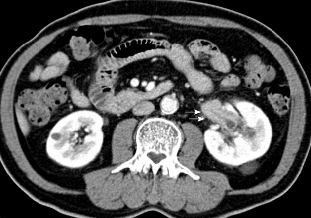
- A 64-year-old man visited our hospital with the chief complaint of microscopic hematuria for the past 2 months. Laboratory tests were unremarkable and urine cytology was normal. Abdominal computed tomography (CT) revealed a homogeneous solid tumor with slightly low attenuation in the left renal sinus (Fig. 1). The left renal pelvis had marked wall thickening with peripelvic soft tissue infiltration. The left proximal ureter and major calyces also had thickened walls with infiltrative features. Altogether, these abdominal CT findings were suggestive of invasive urothelial carcinoma arising in the left pelvis with involvement of renal sinus soft tissue. Nephroureterectomy was performed, and the cut surface of the renal sinus revealed a grayish-white solid tumor of rubbery consistency. The tumor was primarily located in the renal pelvis, but extensions to the renal calyces and proximal ureter were suspicious. The pelvic tumor mass measured 4×2.5 cm and exhibited peripelvic infiltrative features (Fig. 2). However, the mucosal surfaces of the renal pelvis, ureter and calyces were smooth, and there was no evidence of urothelial malignancy. Additionally, the mucosal layers of the renal pelvis, calyces and ureter were intact without abnormal urothelial changes. Microscopically, the tumor occupying the renal sinus contained widely scattered, hyperplastic lymphoid follicles, characteristic of a lymphoproliferative lesion. The follicles varied in size and showed polarized germinal centers; moreover, interfollicular spaces were markedly infiltrated by mature plasma cells, highlighted by CD138 immunostaining (Fig. 3). Immunostaining for κ and λ immunoglobulin light chains indicated that the plasma cells were polyclonal in origin. Immunostaining for human herpes virus 8 (HHV-8) was performed, the results of which were negative. These histological and immunohistochemical findings were compatible with a diagnosis of Castleman's disease of the plasma cell type. Three months after the operation, there was no evidence of disease recurrence on follow-up abdominal CT.
CASE REPORT
- The occurrence of Castleman's disease in the renal sinus is exceedingly rare.1 To the best of our knowledge, only a few cases have been reported.1,3-6 The clinicopathological characteristics of the reported cases, involving six males and one female, are summarized in Table 1. Although Castleman's disease generally shows no gender preference,2 cases involving the renal sinus appeared to show a male predominance. The median age for Castleman's disease is in the fourth decade.2 However, the mean age of the cases involving the renal sinus was 65 years. The main presenting symptoms included abdominal pain, weight loss, anorexia, and microscopic hematuria. Three of the cases were clinically unicentric and four were multicentric. Five cases were of the plasma cell type, one of the hyaline vascular type and one of mixed type. The patient in the present case, a 64-year-old male, presented with symptoms of microscopic hematuria, and his disease was histologically classified as plasma cell type and clinically as unicentric type.
- Although the exact pathophysiology of Castleman's disease is unknown, recent reports suggest that HHV-8 infection may stimulate B lymphocytes to induce interleukin 6 (IL-6) production in the mantle zone; IL-6 overproduction has been shown to be associated with the systemic manifestations of Castleman's disease, especially in cases of multicentric disease.7 In our case, immunostaining for HHV-8 was performed, but the result was negative.
- Preoperative diagnostic imaging methods are not useful in differentiating Castleman's disease arising in the renal sinus from other diseases because of a lack of tumor-specific imaging features. Previously, Nishie et al.1 described the CT and magnetic resonance imaging features of three cases of Castleman's disease involving the renal sinus. They reported that the renal sinus lesions formed in Castleman's disease can appear as homogeneous masses with mild enhancement, such as is often observed in malignant lymphomas. Thus, it is difficult, on the basis of radiological findings, to differentiate Castleman's disease from malignant lymphomas infiltrating the renal sinus. Other differential diagnoses may include invasive urothelial carcinoma, granulomatous diseases, sarcomas, and metastases. Consequently, pathological evaluation is currently the only method of identifying Castleman's disease of the renal sinus.
- Here we have described an extremely rare case of unicentric Castleman's disease of the plasma cell type arising in the renal sinus. Although preoperative diagnosis of Castleman's disease is difficult and accurate diagnosis is only possible by histopathologic evaluation, it should be considered in the differential diagnosis of renal sinus tumors.
DISCUSSION
- 1. Nishie A, Yoshimitsu K, Irie H, et al. Radiologic features of Castleman's disease occupying the renal sinus. AJR Am J Roentgenol 2003; 181: 1037–1040. PMID: 14500225. ArticlePubMed
- 2. Cronin DM, Warnke RA. Castleman disease: an update on classification and the spectrum of associated lesions. Adv Anat Pathol 2009; 16: 236–246. PMID: 19546611. ArticlePubMed
- 3. Ryu JH, Oh JW, Kim KH, et al. Castleman disease misdiagnosed as a neoplasm of the kidney. Korean J Urol 2009; 50: 413–416. Article
- 4. Nagahama K, Higashi K, Sanada S, Nezumi M, Itou H. Multicentric Castleman's disease found by a renal sinus lesion: a case report. Hinyokika Kiyo 2000; 46: 95–99. PMID: 10769797. PubMed
- 5. Nolan RL, Banerjee A, Idikio H. Castleman's disease with vascular encasement and renal sinus involvement. Urol Radiol 1988; 10: 173–175. PMID: 3072749. ArticlePubMed
- 6. Park JB, Hwang JH, Kim H, et al. Castleman disease presenting with jaundice: a case with the multicentric hyaline vascular variant. Korean J Intern Med 2007; 22: 113–117. PMID: 17616028. ArticlePubMedPMC
- 7. Menke DM, Chadbum A, Cesarman E, et al. Analysis of the human herpesvirus 8 (HHV-8) genome and HHV-8 vIL-6 expression in archival cases of castleman disease at low risk for HIV infection. Am J Clin Pathol 2002; 117: 268–275. PMID: 11863223. ArticlePubMed
References
Fig. 1Abdominal computed tomography reveals a homogeneous infiltrative mass in the left renal sinus with relative low attenuation compared to the renal parenchyma (arrows).

Fig. 2Gross findings. The resected kidney contains a 4×2.5 cm gray-whitish infiltrative pelvic mass (arrows) with marked thickening of the walls of the proximal ureter and major calyces.
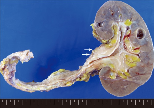
Fig. 3Histopathologic findings. (A) The pelvic tumor is a benign lymphoproliferative lesion with widely scattered, hyperplastic lymphoid follicles. The pelvic urothelium is intact. (B) The renal calyces also contain a submucosal lymphoproliferative lesion. (C) Marked plasma cell infiltration in interfollicular spaces. (D) The plasma cells are positive for CD138 upon immunostaining.
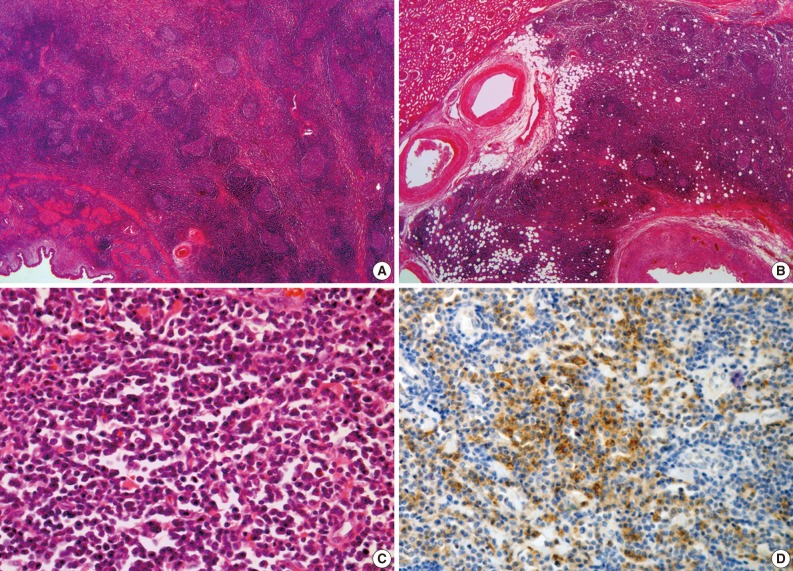
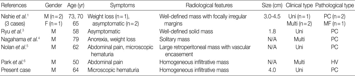
Figure & Data
References
Citations
Citations to this article as recorded by 
- Misdiagnosis of renal pelvic unicentric Castleman disease: a case report
Dian Fu, Bo Yang, Ming Yang, Zhenyu Xu, Wen Cheng, Zhijia Liu, Liming Zhang, Zhiguo Mao, Cheng Xue
Frontiers in Surgery.2023;[Epub] CrossRef - Case report: Castleman’s disease involving the renal sinus resembling renal cell carcinoma
Enlong Zhang, Yuan Li, Ning Lang
Frontiers in Surgery.2022;[Epub] CrossRef - Radiologic features of Castleman’s disease involving the renal sinus: A case report and review of the literature
Xiao-Wan Guo, Xu-Dong Jia, Shan-Shan Shen, Hong Ji, Ying-Min Chen, Qian Du, Shu-Qian Zhang
World Journal of Clinical Cases.2019; 7(8): 1001. CrossRef - Castleman’s Disease: a Suprarenal Surprise!
Praveen Sundar, Priyank Bijalwan, Ginil Kumar Pooleri
Indian Journal of Surgical Oncology.2018; 9(2): 254. CrossRef
 PubReader
 Cite this Article