![]()
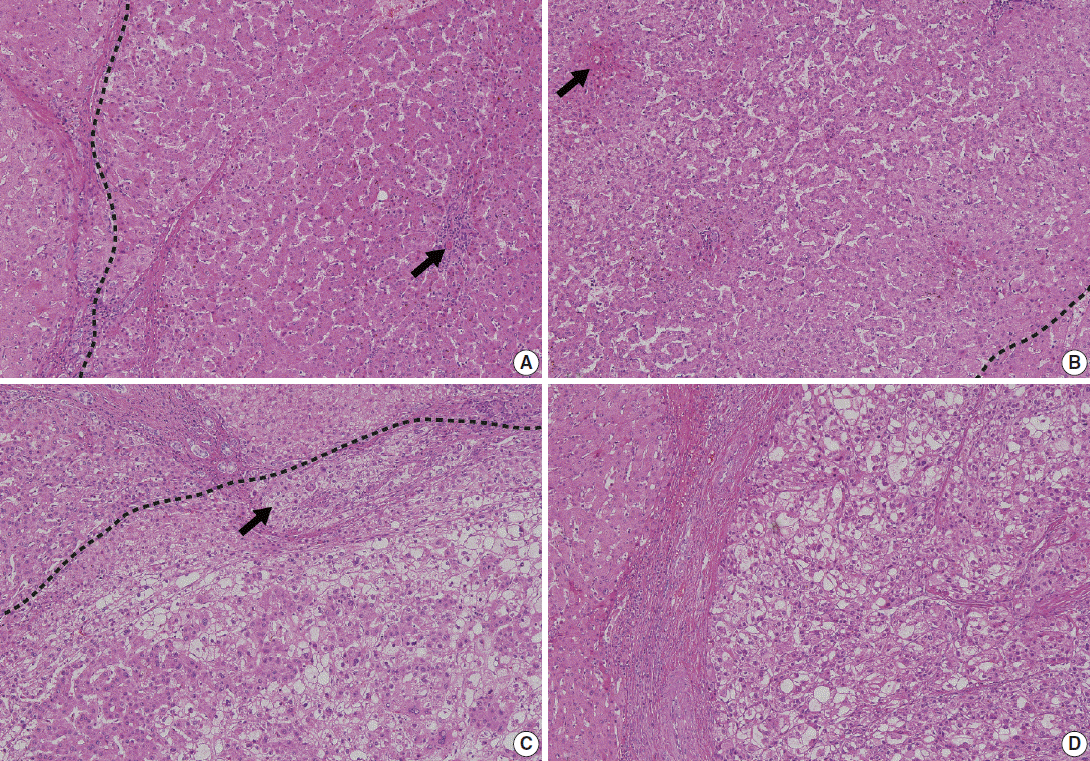
Representative pathologic images of multistep hepatocarcinogenesis. (A) Low-grade dysplastic nodule (right of the dashed line) shows increased cellularity, and residual portal tracts (arrow) are easily identifiable within the nodule. (B) High-grade dysplastic nodule (left of the dashed line) shows further increased cellularity and frequent unpaired arteries (arrow). (C) Early hepatocellular carcinoma (below the dashed line) is poorly demarcated but shows unequivocal cytological atypia and stromal invasion (arrow). (D) Advanced hepatocellular carcinoma is well demarcated by a thick capsule and shows overt features of malignancy.